
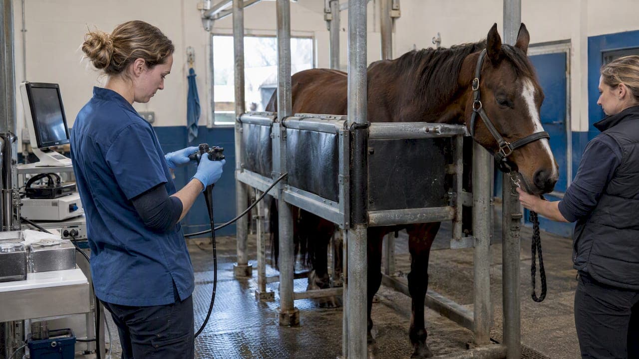
A good equine sinoscopy setup comes down to four things: a scope that fits the sinus and can be sterilized, a trephine kit and a prepared surgical site, a horse that is safely sedated and restrained, and a reprocessing plan ready before the first incision. This guide walks through each in the order you will need them, from choosing the endoscope to cleaning it afterward.
It is written for equine veterinarians and clinic managers who do sinus work in a hospital, a field truck or a barn aisle. It covers the technique at the level of setup and workflow. Sedation protocols, drug choices and case selection stay with the attending veterinarian.
TL;DR
- An 8 mm flexible endoscope is described as ideal for sinoscopy, and a 4 mm arthroscope is another option. A working channel helps with biopsy and sampling.
- Sinoscopy is done standing under sedation, with local anesthetic at the site. Portals are made in the frontal or maxillary bone with a 9.5 mm to 15 mm Galt trephine.
- Plan for hemorrhage and portal wound infection. Have sterile gauze, irrigation and suction ready.
- Brush, flush, soak and dry the scope after every case, following the manufacturer's instructions for the disinfectant.
- Set up the whole tray and the reprocessing station before the horse is sedated, so nothing is improvised with a needle in the head.
Table of Contents
- What sinoscopy is and when you set up for it
- Choosing the endoscope
- The sinoscopy tray
- Room and horse setup
- Step-by-step: from incision to closure
- Complications to plan for
- Cleaning and reprocessing after the case
- A pre-procedure checklist
- Our perspective
- Building your equine kit
- Related reading for equine and small-animal teams
- FAQ
- Sources
What sinoscopy is and when you set up for it
Sinoscopy is direct endoscopic examination of the paranasal sinuses through a small bone portal. It lets you see mucosa, fluid, masses and the region of the cheek teeth roots, and it lets you sample or treat what you find. It is generally done standing under sedation, using either an arthroscope or a sterile flexible fiberoptic or video endoscope, as described in dvm360's review of standing sinoscopy.
Most clinics reach for it after radiographs or a nasal endoscopy exam leave questions: chronic nasal discharge, suspected sinusitis, a mass, or a tooth root problem. The setup below assumes you have already decided to go in.
Choosing the endoscope
The scope is the biggest decision in the setup, and the literature points in a clear direction.
Flexible or rigid
A French veterinary practice review states that an 8 mm flexible endoscope is ideal, with a 4 mm arthroscope as another option. The reasoning is practical. A 2011 BEVA congress paper on IVIS notes that flexible endoscopes offer better diagnostic value than rigid arthroscopes because they can steer around structures inside the sinus and reach areas such as the cheek tooth apices.
A rigid scope still has a place. It is compact, easy to hold steady and a familiar tool for anyone who does joint work. If your practice already owns arthroscopes, they can serve for lighter cases and for portals where a straight view is enough.

Working channel
A working channel changes what you can do through one portal. The same French review notes that a channel makes biopsies and sampling possible, and that a rongeur or diode laser can be used for fenestration. If you expect to take tissue, check that your scope has a channel and that your flexible biopsy forceps fit it before the day of the procedure.
Video, light and recording
Sinoscopy is a team procedure. The surgeon, the person handling the horse and often a student or technician all benefit from a monitor they can see. Choose a system with a bright light source and a screen positioned where the surgeon does not have to twist. Recording to an SD card gives you images for the record and for the owner, and it is simple in the field because there is no separate processor to power.
If you work on the road, a self-contained monitor-type unit makes the setup much lighter. Our 8 mm equine airway inspection field scope with monitor and SD-card recording and the USB version of the 8 mm airway scope are built for that kind of portable work. Whichever scope you choose, confirm with the manufacturer that its insertion tube and sterilization method suit sinus use before you plan the case.
For general guidance on matching scope diameter to the job, see our article on small diameter endoscopes in veterinary practice. For airway-specific equipment, read our equine airway endoscopy equipment guide.

The sinoscopy tray
Build one tray and keep it the same every time. A consistent tray is how a busy practice avoids hunting for a trephine while the horse is sedated.
| Item | Purpose |
|---|---|
| Sterile flexible endoscope or arthroscope | Visualization |
| Sterile sleeve or drape for the scope and light cable | Keeps the field clean |
| Galt trephine, 9.5 mm to 15 mm | Creates the bone portal |
| Scalpel and blade | Skin incision |
| Local anesthetic, syringes, needles | Desensitizing the site |
| Sterile saline, irrigation set, suction | Clearing blood and exudate |
| Biopsy forceps, rongeur or laser if planned | Sampling and fenestration |
| Sterile gauze | Hemorrhage control and packing |
| Staples or suture | Skin closure |
| Foley catheter, if lavage is planned | Post-procedure sinus lavage |
The trephine size range comes from dvm360's standing sinoscopy article, which describes portals made with a 9.5 mm to 15 mm Galt trephine. Match the portal to the scope you plan to pass, and open a second size on the tray if you are unsure.
Protecting the scope on the way in
Bone edges are the quiet killer of insertion tubes. The French review advises protecting the endoscope during insertion, for example by placing the distal sheath through a syringe casing or making sure the bone edges are smooth. Put a sterile syringe casing on the tray as a standard item. A damaged bending section or a nicked outer sheath is an expensive repair and a patient infection risk. If a scope does take damage, our endoscope repair service is where to start.
Room and horse setup
Where the horse stands
Sinoscopy is a standing procedure, so the layout is about the horse's safety and the surgeon's footing. Use stocks if you have them. In a barn aisle, pick a spot with a non-slip floor, room to retreat and a wall or stocks on one side. Keep the monitor in the surgeon's line of sight, not behind the horse's head.
Handling and head position
The University of Minnesota's large animal surgery guide on sinus trephination makes a safety point worth repeating: in a standing horse, the pin and handle can be thrown violently if the horse jerks its head. Assign one person to the head whose only job is the head. Support it, watch the sedation level and warn the surgeon at the first sign of a head toss. The surgeon should keep clear of the line of the handle when the horse is not settled.
Sedation and local anesthesia
Dvm360 reports that sinoscopy is done standing under sedation, and that the surgical site is prepared and desensitized with a subcutaneous injection of local anesthetic. The sedation protocol is the attending veterinarian's call, based on the horse's temperament, health and the planned duration. Have the sedation top-up drawn up and labeled before you start.
Tetanus status
The Minnesota guide recommends tetanus prophylaxis for horses undergoing sinus trephination. Check the vaccination record at intake, not at the stocks, so there is time to give antitoxin or a booster per your veterinarian's plan.
Step-by-step: from incision to closure
The sequence below follows the standing technique described by dvm360. Adjust it to your own protocol and your case.
- Confirm the site. Use your radiographs, and any nasal endoscopy findings, to choose between a frontal and a maxillary portal. Mark the site.
- Prepare and desensitize. Clip and scrub the skin, then infiltrate local anesthetic subcutaneously. Wait for it to take effect before cutting.
- Make the skin incision. Keep it only as long as the trephine needs.
- Create the portal. Use the Galt trephine in the frontal or maxillary bone, per the technique dvm360 describes. Smooth any rough bone edge.
- Insert the scope. Pass the protected, sterile scope through the portal. Initial irrigation or suction may be needed to clear the field.
- Explore systematically. A conchofrontal portal is frequently used to look into several compartments. It is often combined with fenestration of the maxillary septal bulla, which gives access to the rostral maxillary and ventral conchal sinuses, as the same dvm360 source explains.
- Sample or treat. Use the working channel for biopsy, or a rongeur or diode laser for fenestration.
- Withdraw and close. The skin incision is closed with staples or sutures.
Work out the roles before step 1. One person on the head, one on the scope and light, and one on irrigation, suction and the tray keeps the field calm.
Complications to plan for
Planning for complications is part of the setup, because the supplies need to be in the room.
Hemorrhage
Bleeding is the most frequent complication. An Austrian study comparing trephination with a minimally invasive transnasal approach reports that hemorrhage is the most frequent complication of sinoscopy and may require packing the cavity with sterile gauze for about two days. Keep extra sterile gauze on the tray and decide in advance who is calling for it. Suction that works matters more than a good plan once the view turns red.
Portal wound infection
A long-term UK study of sinoscopic treatment found portal wound infection in nine cases, which is nine of 155 in the report. This is small but real. Strict sterile handling of the scope, sleeve and trephine, careful skin closure and a check of the portal at follow-up are the practical defenses.
Alternatives in selected cases
The Austrian study also concludes that minimally invasive transnasal techniques look like a promising alternative to classical extra-nasal approaches in selected cases. That is useful context when you choose the access route, and a flexible scope with a narrow insertion tube is the instrument that keeps those options open.
Cleaning and reprocessing after the case
A sinoscopy scope has been in a bone portal and a sinus that may hold purulent material. Treat reprocessing as part of the procedure, not an afterthought.
A Kansas State University study on the efficacy of endoscope disinfection describes a sequence of brushing and flushing the channels, then immersion in enzymatic cleaner, water, disinfectant and water again, a 70% isopropyl alcohol rinse and drying. In that work the disinfectant contact time was 8 minutes for accelerated hydrogen peroxide or 12 minutes for ortho-phthalaldehyde. The study also explains that high-level disinfection typically means enzymatic cleaning followed by immersion in a disinfectant such as these, according to the manufacturer's guidelines. Always follow your scope maker's instructions for compatible chemicals and contact times, since they decide your warranty and your scope's life.
Our own guides go deeper on each stage:
- Sterilization in veterinary endoscopy covers the methods and when each applies.
- Step-by-step endoscope sterilization is a checklist you can post at the sink.
- How to store endoscopic equipment helps you keep a clean scope clean.
- Prepping an endoscope for inspection is useful for the pre-procedure check.
A simple habit helps: leak-test and inspect the scope before and after each use, and log it. Catching a small sheath nick from a bone edge early is cheaper than finding it after a fluid ingress.
A pre-procedure checklist
Print this and keep it with the tray.
- Radiographs and any prior endoscopy reviewed, portal site chosen
- Tetanus status confirmed
- Scope, sleeve, light and monitor tested, recording media inserted
- Sterile tray open: trephine in the planned size and one alternate, blades, irrigation, suction, gauze, closure material
- Syringe casing or other scope protection available
- Biopsy forceps, rongeur or laser checked if sampling is planned
- Head handler briefed, stocks or safe space set
- Reprocessing station ready with enzymatic cleaner, disinfectant, brushes and fresh water
Our perspective
We see the same pattern in the clinics we supply: the practices that do sinus work well are the ones that treat the scope as a system, not a single instrument. The scope, the light, the monitor, the sleeve, the protected insertion and the reprocessing station all have to work together.
That is why we focus on portable, self-contained equine endoscopes at a lower cost than hospital-grade human systems. A field vet should not need a rack of equipment to look into a horse's sinus, and a small practice should not have to choose between owning a scope and owning a good one. Monitor-type units with SD-card recording keep the setup short, and a working channel with the right biopsy forceps keeps one portal useful for both looking and sampling.
We also believe in keeping scopes working. Bone edges, bending and routine wear are part of sinus work, and our team handles repairs and supports clinics with questions around the clock. If you are building a sinoscopy kit, start with the scope you will actually carry to the barn, and build the tray around it.
Endoscope
Building your equine kit
If you are putting a sinoscopy setup together or replacing an aging scope, we carry the pieces in one place. Our equine 8 mm airway field scope with monitor suits clinics that work on the road, and our semi-portable 3 meter equine endoscope is an option when you also need length for longer exams. Add flexible biopsy forceps and rigid scopes and arthroscopes to cover sampling and straight-view work. Browse the full range at 1800endoscope.com.
Prices and availability change, so check each product page for the current details, and contact us if you want help matching a scope to your caseload.
Related reading for equine and small-animal teams
Sinus work often goes hand in hand with dental and airway exams. Our article on the equine dental endoscope covers oral examination, which is useful when a cheek tooth is the suspected source. If your practice also sees dogs and cats, the feline rhinoscopy setup guide and small animal endoscopy tips show how the same habits of preparation and cleaning carry over.
FAQ
What size endoscope do you need for equine sinoscopy?
An 8 mm flexible endoscope is described as ideal, and a 4 mm arthroscope is another option. Flexible scopes are favored because they can steer around structures inside the sinus. Match the scope to the portal size you plan to create with the trephine.
Can sinoscopy be done on a standing horse?
Yes. It is commonly performed standing under sedation, with local anesthetic infiltrated at the surgical site. Good head control and a clear plan for sedation top-ups are the main safety measures.
What are the most common complications?
Hemorrhage is the most frequent, and it may call for packing the cavity with sterile gauze for about two days. Portal wound infection is also reported, in nine of 155 cases in one UK study. Sterile technique and follow-up checks of the portal reduce the risk.
How do you clean the endoscope after sinoscopy?
Brush and flush the channels, then soak in enzymatic cleaner, water, high-level disinfectant and water again. Rinse with 70% isopropyl alcohol and dry. Follow the scope manufacturer's instructions for the disinfectant and contact time.
Sources
- Sinoscopy in the standing horse (dvm360)
- Sinusoscopie chez le cheval : intérêt diagnostique et thérapeutique (Le Point Vétérinaire)
- Sinoscopy (IVIS, BEVA Congress 2011)
- How to: Sinus trephination (University of Minnesota, Large Animal Surgery)
- Trephination versus minimally invasive transnasal approach (PMC)
- A long-term study of sinoscopic treatment of equine sinus disease (Equine Veterinary Journal)
- Efficacy of disinfection of endoscopes (Kansas State University)