
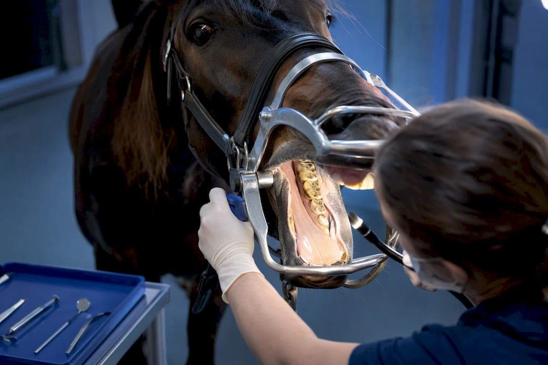
An equine dental endoscope is a rigid intraoral imaging system that markedly increases visualization and documentation compared to a mirror exam. It belongs in a modern equine dentistry toolkit whenever a practice needs better detection, clearer client education, and a permanent record of findings. It complements radiography rather than replacing it, since apical disease, sinus pathology, and EOTRH often need imaging that endoscopy cannot provide on its own.
TL;DR:
- Endoscopy offers significantly higher sensitivity than mirror exams for detecting surface lesions, but it can produce more false positives, requiring experienced interpretation.
- Choosing a system should prioritize workflow, durability, and support, with smaller diameters and better illumination being more important than pixel count or 4K resolution.
- Workflow practices like thorough rinsing and consistent file naming enhance image quality and diagnostic efficiency, often outweighing equipment specifications.
- Endoscopy functions best as a monitoring tool for borderline issues and complements radiography; it does not replace imaging for deep or sinus-related pathologies.
- Vendors should be evaluated based on support, repair turnaround, and sample footage, with full system costs including accessories often exceeding the scope's initial price.
Table of Contents
- What Is an Equine Dental Endoscope Used For?
- What Components and Specs Matter Most?
- Is an Endoscope More Accurate Than a Mirror Exam?
- How Do You Run an Endoscopic Dental Exam Step by Step?
- How Do You Choose the Right System for Your Practice?
- What Should You Verify Before You Buy?
- Why Most Practices Underuse What Endoscopy Actually Offers
- Where to Find an Equine Dental Endoscope System
- Sources
- FAQ
What Is an Equine Dental Endoscope Used For?
Two device categories dominate equine practice: rigid oral endoscopes built specifically for the caudal mouth, and handheld intraoral cameras adapted from general veterinary or human dental use. Rigid systems tend to give better caudal reach and a wider viewing angle, which matters once you're past the premolars.
Clinically, an endoscope earns its keep in four places: initial inspection of teeth and soft tissue that a mirror can't angle into, documentation for the medical record or referral letter, intraoperative guidance during extractions or periodontal work, and client or student teaching, since a monitor lets everyone see the same lesion at once. It also supports monitoring progression of borderline findings, such as a small periodontal pocket or minor infundibular defect, over successive visits. None of this displaces the mirror exam or radiography. Think of endoscopy as the layer that catches what a mirror misses and gives radiography a visual target to confirm.

What Components and Specs Matter Most?
A working system is really five parts: the scope head, the shaft, the light source, the camera and monitor, and the sheath that protects it all between patients. Each spec has a direct clinical consequence.
- Shaft diameter: typically 5 to 15 mm; smaller diameters improve patient comfort and maneuverability around the bit and speculum bars, per IVIS/AAEP proceedings on incorporating oral endoscopy.
- Working length: roughly 50 cm is standard, enough to reach the caudal cheek teeth without repositioning the horse's head repeatedly.
- Viewing angle: ranges from 60 to 115 degrees; wider angles help visualize occlusal and interproximal surfaces on the last molars.
- Illumination: LED heads built into the scope avoid a separate fiber-optic light cable, though external light sources still deliver more consistent brightness for video recording.
- Camera resolution and monitor/recording options: matter less than most buyers assume once you clear a basic HD threshold.
- Protective sheaths: extend scope life and simplify disinfection between patients.
Connectivity ranges from wired camera towers to USB or SD card recorders and wireless streaming to a tablet. Wireless setups suit ambulatory work where hauling a monitor cart isn't practical, but they add a battery and signal dependency that wired systems don't have.
Pro Tip: Don't chase pixel count. A crisp 1080p image with good illumination and a stable shaft beats a 4K sensor paired with a dim, wobbly light source every time.
Is an Endoscope More Accurate Than a Mirror Exam?
Yes, and the evidence is more specific than "endoscopy is better." A 2022 blinded prospective study compared both methods against post-mortem examination and found oral endoscopy achieved 83% sensitivity versus 39% for mirror exam.
Endoscopy caught more real lesions but also generated more false positives than the mirror, and the study's authors flagged their small sample size as a real limitation on how far these numbers generalize.
That trade-off matters at the chairside. Endoscopy is the better screening tool for catching subtle periodontal pockets, early infundibular decay, or caudal lesions a mirror simply cannot angle into. But a positive finding on endoscopy still benefits from experienced interpretation before it drives a treatment decision, and it won't answer every diagnostic question. Apical infection, sinus disease, and EOTRH typically require radiography or CT, because those pathologies live below or behind the surface an endoscope can actually see.
How Do You Run an Endoscopic Dental Exam Step by Step?
Preparation determines image quality more than any spec on the scope itself. Field workflow, not camera resolution, is what separates a usable exam video from a frustrating one.
- Sedate appropriately for the procedure and place a full-mouth speculum, following standard AAEP-aligned technique.
- Position the head at a comfortable working height and rinse the oral cavity thoroughly to clear feed debris before inserting the scope.
- Start rostral at the incisors and canines, then move caudally through each cheek tooth in a consistent sequence so nothing gets skipped.
- Angle the tip to inspect occlusal surfaces, interproximal spaces, and the gingival margin, using the wide-angle lens to catch the last molars.
- Capture stills at each notable finding and switch to video only when documenting motion, like probing a periodontal pocket or an intraoperative step.
Name files by date and tooth number, not just patient name. It saves real time when you're comparing images across visits.
Pro Tip: Rinse twice, not once. A second rinse after speculum placement clears the debris that settles once the horse starts salivating.

How Do You Choose the Right System for Your Practice?
Start with how and where you'll use it. Ambulatory practitioners need battery life, a compact case, and a scope tough enough to survive a truck bed; in-hospital setups can prioritize a larger monitor and a stationary camera tower instead.
Evaluate the whole system, not just the scope head. That means the light source, camera and control unit, recording method, sheaths, and your clinic's cleaning and disinfection pathway between patients. A modular, cost-conscious build can work well for a solo practitioner who wants to add components over time rather than buying everything at once.
Before you sign anything, ask vendors for:
- Sample video and still images captured in an actual clinical setting, not a marketing reel
- A written warranty and repair turnaround time
- Spare-part lead times, especially for the shaft and light cable, which take the most wear
- Cleaning and disinfection instructions compatible with your clinic's protocols
- Service-level agreements covering loaner equipment during repairs
Price varies widely by scope diameter, monitor type, and recording features, so budget for the full system rather than anchoring on the cheapest scope head you find. Recurring costs, sheaths, cable repairs, occasional recalibration, tend to matter more over three years than the sticker price on day one.
What Should You Verify Before You Buy?
Vendor support is where good systems turn into reliable ones. Before committing, confirm parts availability, typical repair turnaround, whether cleaning instructions are clearly documented, and what training resources come with the purchase.
1800endoscope focuses specifically on veterinary and industrial endoscopy, with product pages and technical guides that lay out these details rather than leaving them to a sales call. Treat any vendor's claims, including 1800endoscope's, as something to verify: request demo footage shot on an actual horse, ask for a reference clinic using the same model, and get the warranty and repair policy in writing before the invoice arrives. The AAEP position on equine dentistry is worth reviewing too, since it frames how endoscopic findings should integrate into diagnosis and treatment planning rather than stand alone.
Why Most Practices Underuse What Endoscopy Actually Offers
The conventional pitch for endoscopy focuses almost entirely on catching more lesions, and the sensitivity numbers back that up. But the more useful shift in thinking is treating endoscopy as a monitoring tool, not just a detection tool. A borderline periodontal pocket or early infundibular defect doesn't always need immediate treatment. It needs a baseline image and a follow-up comparison six or twelve months later, and that's exactly what endoscopic documentation is built for.

Where most buying decisions go wrong is chasing camera specs instead of workflow. A clinic that standardizes speculum placement, rinsing, and file naming will get more diagnostic value from a modest system than a clinic with a premium scope and no consistent protocol.
If you're prioritizing one thing first, prioritize the full system, cleaning pathway, recording workflow, and service support, over the marginal difference between two similarly specced scope heads. That decision will affect your daily workflow far longer than any spec sheet comparison.
— Endoscope
Where to Find an Equine Dental Endoscope System
Once you know the specs and workflow you need, the next step is matching them to an actual system. 1800endoscope carries rigid and portable endoscopy equipment built for veterinary use, along with the accessories that round out a complete setup: sheaths, light sources, and recording components.

For clinics comparing diameter and reach, the equine field scope with monitor-type SD recording is a practical entry point for ambulatory work, and it's worth requesting sample video before you commit to a specific model. Some vendors also handle endoscope repairs directly, which matters when a caudal shaft takes the kind of wear ambulatory practice guarantees. Reach out for a datasheet or ask about service agreements before your next equipment budget cycle closes.
Sources
- Blinded comparison of mirror and endoscopic oral examination in the horse: Sensitivity, specificity and observer agreement
- How to Incorporate Oral Endoscopy Into an Equine Dental Examination (IVIS/AAEP proceedings)
- Position on Equine Dentistry – AAEP
FAQ
How Much Does an Endoscopy Cost for a Horse?
Cost depends on whether the exam uses a clinic's existing equipment or requires a referral to a specialist with advanced imaging, along with regional pricing and sedation needs. Current equipment pricing for endoscope systems is listed directly on the 1800endoscope site rather than a fixed number, since configurations vary by scope diameter and accessories.
How Much Does a Periodontal Endoscope Cost?
Periodontal-capable endoscope systems vary by shaft diameter, viewing angle, and whether recording and light source components are bundled or purchased separately. Rather than a single flat figure, practices should request a quote covering the full system, scope, light source, camera, and sheaths, since that combination drives most of the cost.
How Much Does a Horse Dentist Visit Cost?
A routine equine dental visit typically covers sedation, a full oral exam, and floating, with cost varying by region and whether advanced imaging like endoscopy or radiography is included. Practices offering endoscopic documentation as part of the exam may charge more than a basic floating appointment, reflecting the added diagnostic value.
Is EOTRH in Horses Painful?
Yes, EOTRH (equine odontoclastic tooth resorption and hypercementosis) is a progressive, painful condition affecting the incisors and canines, typically in older horses. It often requires radiography to confirm, since full-mouth imaging detects EOTRH cases not evident on oral exam, particularly in older horses.
Does an Endoscope Replace the Need for Dental X-Rays?
No. Endoscopy improves visualization of surface and near-surface lesions, but it cannot see apical infection, sinus disease, or bone changes below the tooth structure. The AAEP position on equine dentistry treats endoscopic findings as one input among several, not a substitute for radiography when deeper pathology is suspected.