
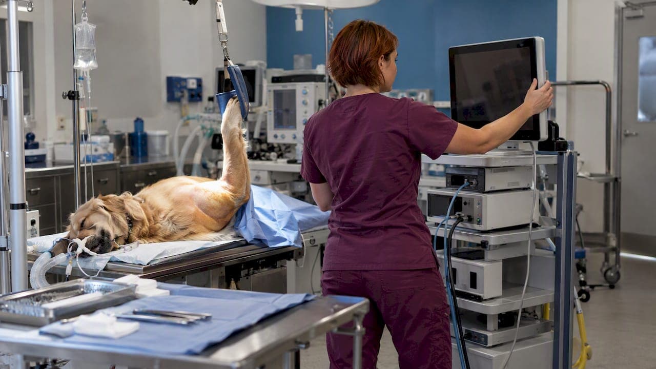
A working veterinary arthroscopy setup has five parts: a small-diameter rigid scope with a cannula, a light source and cable, a camera with a good monitor, a fluid system, and a prepared room and patient. Get any one of them wrong and the picture suffers, because the image can only be as good as the weakest link in the chain. This guide walks through each part in the order you build it, then covers portals by joint, handling and sterilization, and after-care.
TL;DR
- A diagnostic system needs a 30° short arthroscope (1.9, 2.4 or 2.7 mm depending on patient and joint), cannula and blunt trocar, light source and cable, camera, monitor, fluid bags and sterile tubing, and a way to document images.
- Treat scope, light cable, camera and monitor as one imaging chain. Upgrading one link will not help if another is the bottleneck.
- Use lactated Ringer's, keep pressure low enough to avoid extravasation, and choose gravity or pump delivery on purpose.
- Portal placement depends on joint and procedure, so plan the monitor position, limb positioning and extra instruments before the dog is draped.
- Sterilize only by methods the manufacturer approves, and rinse glutaraldehyde thoroughly.
Table of Contents
- The imaging chain: what you need on the cart
- Fluid management: gravity or pump
- Instruments and accessories
- Room layout and patient preparation
- Portals and triangulation by joint
- The procedure in sequence
- Handling and sterilization
- After-care and monitoring
- Perspective: what we see in clinics buying their first system
- Where our equipment fits
- FAQ
- Sources
The imaging chain: what you need on the cart
Veterinary arthroscopy borrows from human orthopedics, but the patient range runs from a 5 kg dog to a horse or a calf. That is why the scope itself is the first decision.
A diagnostic system described in the WSAVA 2015 Congress proceedings includes:
- an arthroscopic camera with a quality color monitor
- a short, 30° arthroscope of 1.9, 2.4 or 2.7 mm, chosen for joint and patient size
- a compatible cannula and blunt trocar
- a light source
- lactated Ringer's bags and sterile tubing
- a photo-documentation system
An earlier WSAVA basic-principles paper adds a fiber-optic light cable, a 13 to 19 inch HD monitor, a fluid infusion set and basic instruments. It calls videoarthroscopy the only acceptable method, because it gives magnification, lets the whole team see, and makes surgery practical.
Scope size and the cannula
The arthroscope is used inside a cannula or sheath. The sheath protects the telescope and gives fluid a channel in. That means the scope, cannula and trocar must be sized as a matched set. The scrub nurse should check this before the case starts, not when the first portal is being made.
For smaller joints such as the canine elbow, a 2.4 mm scope is a common starting point. We go deeper on that choice in our article on 2.4 mm canine scope size for elbow cartilage injury. Larger dogs and the stifle or shoulder often tolerate a 2.7 mm scope, while 1.9 mm suits the smallest patients and tight joints.
Light source, camera and monitor
The light cable carries light to the scope, and the camera head clips onto the eyepiece. Compatibility needs a check. The camera control box is specific to the camera, and some light-source connections are manufacturer-specific, although many fit almost any fiber-optic light cable. Buying a mixed system without confirming adapters is the most common way to end up with a cart that does not work on day one.
Monitor placement matters as much as monitor size. Put it where the surgeon looks naturally while the hands work, which is usually directly opposite the surgeon or dorsal to the patient for a shoulder.
Recording and documentation
Photo and video capture are part of the system, not an extra. They support the medical record, client communication and teaching. Our guide on optimizing endoscope video recording for vets covers settings and storage, and our piece on video imaging in animal diagnosis explains why image quality matters clinically.
Fluid management: gravity or pump
Fluid is what makes arthroscopy possible. Distension and continuous irrigation increase working room, decrease bleeding and allow inspection. Lactated Ringer's is the fluid of choice, and pressure is typically held at 40 to 150 mm Hg, below the level that causes pericapsular extravasation.
You have two ways to deliver it:
| Method | How it works | Where it fits |
|---|---|---|
| Gravity-fed bags | Bags hung at height push fluid through sterile tubing | Simple, low-cost setups, small joints, diagnostic cases |
| Pressure-controlled pump | Pump holds a set pressure through the case | Longer procedures, joints that bleed or cloud easily |
Both are described in the WSAVA 2015 material as ways to keep the visual field clear. Pumps are not automatically better. In a human knee study with a tourniquet, automated pumps did not reduce bleeding, air bubbles or loose fibrous tissue compared with gravity, though they did reduce turbidity. That is a human trial, so treat it as a hint rather than a rule for dogs.
The same WSAVA source warns that excessive pressure, particularly from volume-controlled pumps, risks extravasation into surrounding tissues. Whichever method you choose, watch the tissue around the joint for swelling during the case.
Fogging and condensation can also ruin an otherwise good setup. We cover practical fixes in endoscope fogging prevention.
Instruments and accessories
A diagnostic scope alone will not finish most cases. Typical additions include a probe, small grasping forceps, biopsy forceps, and for treatment a motorized shaver or other power equipment. Our overview of veterinary scoping accessories explains what to stock, and flexible biopsy forceps are on our site if your protocol includes sampling.
Keep a spare cannula, a spare light cable and spare fluid sets on hand. A failed light cable mid-case cancels the procedure faster than any other single item.
Room layout and patient preparation
Who does what
The WSAVA 2015 paper splits the assistants' roles clearly. The OR nurse knows the startup sequence and connects cables to the right camera and light boxes. The scrub nurse cleans film from the camera and arthroscope glass with sterile, moistened soft cotton or manufacturer wipes, then confirms that trocar, cannula and scope sizes match. Writing this down as a checklist removes most setup errors.
Anesthesia and aseptic prep
Arthroscopy in dogs is done under general anesthesia after routine clipping and scrubbing, as laid out in the WSAVA basic-principles paper. The American College of Veterinary Surgeons likewise describes general anesthesia and aseptic preparation as typical for equine arthroscopy. Clip widely enough that you can extend a portal or convert to an open approach without re-prepping. Plan the anesthesia protocol with your anesthetist before the day, including a contingency plan, as human surgical guidance on knee arthroscopy room set-up also advises.
Positioning example: canine shoulder
For the canine shoulder, Bardet's chapter describes lateral recumbency with the affected shoulder uppermost, the monitor dorsal to the patient and the limb suspended. The lateral portal is the usual telescope portal and the caudolateral portal the usual instrument portal. Egress is optional.
Portals and triangulation by joint
Portal location varies with both joint and procedure. The basic technique is triangulation: the scope goes through one portal, instruments through another, and extra portals are added for irrigation, access or specialized treatment, per the WSAVA basic-principles page.
Stifle
In the WSAVA stifle technique, the traditional telescope portal sits lateral to the patellar tendon, midway between the tibial tubercle and the patella. The instrument portal is at the same level medially, and the egress portal is superomedial. A motorized shaver is commonly used to open a viewing window through inflamed infrapatellar fat pad before you examine the cruciate ligaments and menisci. That means the shaver and its tubing belong on the cart for any stifle case.
Hock
Tight joints may need help. A 2014 Journal of Small Animal Practice study found that distraction can increase the available tibiotarsal joint space in dogs, and venting the hock increased it further. This is potentially useful for maneuvering instruments inside the joint.
Elbow: respect the ulnar nerve
A 2025 canine cadaver study found that a caudomedial elbow portal placed near the ulnar nerve caused direct nerve injury in 16 of 30 dogs (53%), and injury or high risk of trauma in 21 of 30 (70%). The authors conclude that this portal should be used only if the nerve's course can be precisely determined. Landmark carefully and consider an alternative approach.
Horses and cattle
Large-animal setups change the scope and the positioning, not the principles.
- Equine temporomandibular joint: a 2022 study describes either a flexible needle arthroscope or a 2.5 mm, 30° standard system, with caudal access and saline distension. In standing horses, sedation, local analgesia and a modified mouth speculum helped prevent jaw motion and allowed access to the compartments.
- Bovine stifle: a peer-reviewed bovine study found femoropatellar arthroscopy was best accessed laterally between the middle and lateral patellar ligaments. Different medial and lateral approaches were needed to see the axial and abaxial parts of the femorotibial compartments.
The procedure in sequence
Once the room is ready, the procedure follows a predictable order, summarized from the WSAVA basic-principles paper:
- Anesthetize, clip and scrub, and drape.
- Distend the joint with fluid.
- Insert the cannula and arthroscope, and establish irrigation.
- Explore the joint by triangulation, adding cannulas for irrigation or treatment as needed.
- Lavage thoroughly and close the skin.
Before step 1, run a pre-flight check on the cart: white balance the camera, confirm the image on the monitor, check the light output, and confirm the fluid line flows without air.
Handling and sterilization
Arthroscopes are precise and fragile. Per Veterian Key, handle the scope, camera and light cable carefully, and sterilize them only by manufacturer-approved methods. Those can include ethylene oxide, peracetic acid, hydrogen peroxide gas plasma, or steam only if the device is rated for it. If you use glutaraldehyde, rinse thoroughly. Check your own device's instructions for use, because ratings differ between scopes.
Do not coil light cables tightly, store scopes in protective cases, and never force a scope into a cannula. Scopes that are damaged in handling are a common reason for repair, and our endoscope repair service exists for exactly that.

After-care and monitoring
After-care is set by the surgeon for the individual case, so the following is general. Specialty surgery practices describe arthroscopy as less invasive than open surgery, with most pets leaving the hospital within 24 hours, pain controlled with medication and vigorous activity such as running and jumping discouraged during healing. Short, controlled walks are typically allowed. In your clinic, build the plan around the procedure performed: pain management, incision checks, and a clear instruction to owners about when to call you for swelling, discharge or worsening lameness. Large animals need their own protocol from the attending surgeon.
Perspective: what we see in clinics buying their first system

We sell veterinary and industrial endoscopes and accessories, and we repair scopes. The pattern we see is that clinics spend heavily on one component and ignore the rest. A clinic buys a premium camera and pairs it with a small monitor, or buys a good scope and then fights fogging and poor light.
Our advice is to specify the whole chain at once, write the startup checklist before the first case, and size the scope to the smallest patient you realistically expect to treat. Veterinary-specific equipment priced for animal practice, rather than hospital-grade human systems, is what keeps arthroscopy within reach of a general or referral clinic. We also think a clinic should pick a supplier it can call when something fails mid-schedule, which is why we offer 24/7 support and repairs.
— Endoscope
Where our equipment fits
Our rigid scopes, arthroscopes and otoscopes page lists the small-diameter rigid scopes for joint work. For flexible diagnostic work, light sources and accessories, see our veterinary endoscopy equipment and endoscopic accessories. Tell us your patient range and joints, and we can help you match scope, light source and cable so the chain works together.
FAQ
What is the minimum equipment for veterinary arthroscopy?
You need a short 30° arthroscope (1.9, 2.4 or 2.7 mm), a matched cannula and blunt trocar, a light source and cable, a camera, a monitor, sterile fluid and tubing, and a way to record images. Basic instruments are added for the procedure. Everything must be compatible with the camera control box and light connection.
Should I use a fluid pump or gravity bags?
Either can keep the field clear. Gravity bags are simple and inexpensive, while pressure-controlled pumps hold steady pressure on longer cases. Whichever you pick, keep pressure below the level that causes extravasation, and watch the tissue around the joint.
Which scope size is right for dogs?
It depends on the joint and the dog. The 1.9, 2.4 and 2.7 mm sizes cover most small animal work, with smaller scopes for small patients and tight joints. Choose the smallest size you will regularly need and confirm the cannula matches.
How do I sterilize an arthroscope?
Use only methods the manufacturer approves, such as ethylene oxide, peracetic acid or hydrogen peroxide gas plasma, and steam only if the scope is rated for it. Rinse glutaraldehyde off thoroughly. The instructions for use for your model are the final word.
Sources
- How Can I Help in Arthroscopy? WSAVA 2015 Congress, VIN
- Arthroscopy in Dogs: Basic Principles, WSAVA 2001, VIN
- Veterinary Arthroscopy for the Small Animal Practitioner, introduction
- Veterinary Arthroscopy for the Small Animal Practitioner, shoulder chapter
- Arthroscopy, Veterian Key
- Comparison of two arthroscopic pump systems
- Arthroscopy of the Stifle, WSAVA/VIN
- Evaluation of a joint distractor to facilitate arthroscopy
- The Equine Temporomandibular Joint
- Normal arthroscopic anatomy of the bovine stifle
- Risk of Ulnar Nerve Injury Following Caudo-Medial Elbow Portal
- Equine Arthroscopy, ACVS
- Knee Arthroscopy: Patient Positioning, Room Set-Up
- All About Arthroscopy for Pets, OVRS