
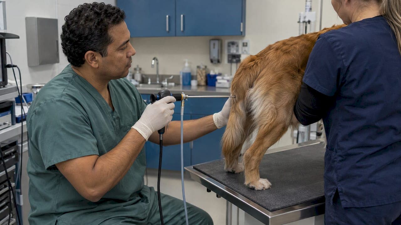
A working vaginoscopy setup for small animal practice is a rigid scope with a protective sheath, a compatible light source and cable, and a way to distend the vagina and pass instruments. A camera and monitor are a worthwhile upgrade. Scope length and diameter should match the size of your patients. Between patients, the scope needs high-level disinfection with a product made for endoscopes, not a general surface disinfectant.
This guide is for clinics and breeding practices choosing or upgrading equipment. It covers each component, rigid versus flexible, video versus direct viewing, sizing, clinical uses, technique basics and reprocessing.
TL;DR
- Rigid scopes are the standard choice for the bitch. A sheath with a working channel allows catheters and brushes to be passed, which a flexible scope handles less easily (Lévy, 2016).
- Plan for at least 30 cm of length for medium and large bitches, a small diameter for small and medium breeds, and an optical angle in the 6° to 30° range.
- Every scope must be matched to a compatible light source. Smaller scopes need brighter light, and worn cables or damaged fibers cost you image quality.
- Reprocess heat-sensitive scopes after every patient with a disinfectant labeled for endoscopes.
- A video camera and monitor make the exam easier to teach, record and show to clients, but the optics are what you buy first.
Table of Contents
- What a vaginoscopy system includes
- Rigid or flexible: which scope for vaginoscopy
- Choosing length, diameter and viewing angle
- Light source and cable compatibility
- When clinics use vaginoscopy
- Preparing the patient and the equipment
- Cleaning and disinfection between patients
- Our perspective
- Build your setup with us
- FAQ
- Sources
What a vaginoscopy system includes
Vaginoscopy lets you look directly at the vestibule, vagina and, with the right scope, the cervix. The equipment is modest compared with a full GI endoscopy tower, and most of it overlaps with what a clinic already uses for urethrocystoscopy or rhinoscopy.
A typical setup has these parts:
- A rigid telescope. Usually a small-diameter scope with a forward-oblique or near-straight view. This is the component that determines what you can see.
- A sheath and obturator. The sheath protects the telescope and carries the irrigation or insufflation channel. The blunt obturator makes entry through the vulva and into the vagina safer.
- A working channel or bridge. This lets you pass a catheter, a brush, a culture swab or small biopsy forceps without withdrawing the scope.
- A light source and light cable. Cold light delivered through a fiber cable.
- A way to distend the vagina. Air from an insufflation bulb or controlled fluid, depending on your technique and scope.
- Optional: camera, monitor and recording. Useful for teaching, record keeping and client communication.
Some clinics start with an otoscope cone or a pediatric-style speculum for a quick look at the caudal vagina. That approach works for basic inspection and for the cytology workflow described in a Clinicians Brief step-by-step guide, but it does not give you the length, magnification or instrument access of a dedicated rigid scope.
Rigid or flexible: which scope for vaginoscopy
In the bitch, a rigid endoscope with a catheter port, or a sheath with a working channel, is better suited to vaginal examination than a flexible one. The reason is practical: the rigid shaft lets you steer within the vaginal canal and pass flexible catheters and brushes through the channel (Lévy, 2016).
That matches how most small animal clinics already work. A rigid scope is also easier to clean and has no articulating section to wear out. Flexible videoscopes remain valuable in other parts of your practice, such as airway or GI work, but for the canine vagina the rigid design fits the anatomy and the task.
Direct viewing versus video
Direct-view telescopes send the image to the eyepiece. Video systems put a camera on the eyepiece or at the scope tip and show the image on a monitor. The trade-offs look like this:
| Factor | Direct view through eyepiece | Video with camera and monitor |
|---|---|---|
| Who sees the exam | One person at a time | The whole team and the client |
| Teaching and training | Harder | Much easier |
| Records | Notes only | Stills and video clips |
| Handling | Lightest setup | Extra cable, camera and monitor to manage |
| Starting investment | Lower | Higher |
If budget forces a choice, buy the right optics first and add the camera later. A good rigid telescope with a camera coupler gives you a path to video without replacing the scope.
Choosing length, diameter and viewing angle
Scope specifications matter more than brand names. Three numbers decide whether a scope works for your patient mix.
Length. To reach the cervix in medium to large bitches, the endoscope should be at least 30 cm long. A shorter scope may show the vestibule and caudal vagina but stop short of the cranial vagina and cervix (Lévy, 2016).
Diameter. The scope must be slim enough to pass the cranial vagina in small and medium breeds. A scope that fits a Labrador will not necessarily fit a Yorkshire Terrier, and forcing it risks trauma. Our guides to choosing the right endoscope diameter and small animal endoscope size walk through how diameter relates to patient size and working channel capacity.
Viewing angle. Most authors recommend an optical angle between 6° and 30° so the cervical opening can be seen as the scope approaches (Lévy, 2016). A slight offset angle helps you look at the dorsal wall and cervical os without bending the shaft against the vaginal wall.

Building a two-scope kit
Clinics that see a mix of toy breeds and giant breeds often end up with two rigid scopes: a short, very slim one for small patients and a longer one for large bitches. If you can only buy one, choose the size that covers your most common patient and confirm the working channel passes the catheters you use. Our small diameter endoscope overview explains where slim scopes fit in a small animal practice.
When you compare options, check these points against the catalog description:
- Working length of the telescope and sheath
- Outer diameter of the sheath, not just the telescope
- Viewing angle in degrees
- Channel size and the catheters or instruments it accepts
- Connector type for the light cable
- Whether the scope can be coupled to a camera
You can browse rigid scope options in our veterinary rigid endoscopy catalog.
Light source and cable compatibility
The light source is easy to overlook and easy to get wrong. Each vaginoscope has to be connected to a light source that suits it, and smaller-diameter scopes demand more luminosity than larger ones because their fibers carry less light. A worn light cable or broken fibers in the scope reduce brightness and limit what you can see (Lévy, 2016).
In practice:
- Match the light post connector and cable adapter to the scope before you order.
- Choose a source with enough output for your slimmest scope, not your thickest.
- Inspect cables regularly. Dark spots when you hold one end to a light suggest broken fibers.
- Keep a spare cable if vaginoscopy is a scheduled service in your practice.
If an image looks dim or uneven, our videoscope troubleshooting guide covers the usual causes before you assume the scope needs service. When it does, we also offer endoscope repairs through our main site.
When clinics use vaginoscopy
Vaginoscopy is a core part of the workup for vulvar and vestibulovaginal disease. The Merck-affiliated MSD Veterinary Manual describes it as the most thorough evaluation of the vestibulovaginal canal, and notes it can usually be done in an estrual bitch during proestrus (MSD Veterinary Manual).
Common reasons to reach for the scope include:
- Abnormal discharge, excessive vulvar licking, perivulvar dermatitis, dysuria, urinary incontinence or breeding difficulty. These can point to urogenital anatomical abnormalities that a scope can confirm or rule out.
- Vaginal lesions. Inspection, digital palpation and vaginoscopy together give the preliminary identification of most vaginal lesions, and surgical treatment is often easier with an episiotomy (IVIS, Current Techniques in Small Animal Surgery).
- Reproductive timing. Vaginoscopic findings such as the color, edema and moisture of the vaginal mucosa add to cytology when evaluating the estrous cycle, including abnormal patterns like early or late ovulation, silent heats or split heats (Reckers et al., Frontiers in Veterinary Science, 2022).
A scope that also fits urethrocystoscopy or rhinoscopy spreads its cost across several services, which is one reason many small animal clinics prefer a slim, versatile rigid scope.
Preparing the patient and the equipment
A short checklist before each exam saves time and protects the scope.
- Check the light path. Connect the cable, switch on the source and confirm a bright, even image on a white surface.
- Assemble the sheath. Insert the telescope, confirm it locks, and fit the obturator if you are entering blind.
- Lay out your instruments. Catheter, culture swab, brush or biopsy forceps within reach. Our flexible biopsy forceps page lists accessory options for the channel.
- Prepare insufflation. Have your air bulb or fluid line ready and tested.
- Plan restraint. Decide before you start whether the patient will stand or need sedation.
Sedation and restraint
Vaginal exploration is generally well tolerated without sedation in standing bitches. Sedation or full anesthesia can be considered in anestrus, with a narrow lumen, in a nervous bitch, or when vestibulitis or vaginitis is present, to prevent trauma or damage to the equipment. The cited paper gives medetomidine at 10 to 20 μg/kg IV as an example (Lévy, 2016). Dose and drug choice are the attending veterinarian's decision for the individual patient.
Clinicians Brief notes that most dogs in proestrus or estrus cooperate, while mild sedation to full anesthesia may be needed in diestrus or anestrus, in anxious dogs, or when pathology may cause pain (Clinicians Brief, 2024).
Basic technique
For a simple otoscope-cone exam, the same source describes moistening the cone with saline or sterile water, introducing it at the dorsal commissure of the vulva, and advancing it dorsocranially at about 45 degrees to avoid the clitoral fossa, until the vagina levels out toward the cranial vagina. A rigid scope follows the same path of least resistance.
Advance gently. Complications of vaginal endoscopy are rare and mostly come from introduction of the scope. If the dog shows defensive movements, stop the exam until restraint is better, which prevents trauma or perforation of the vaginal wall (Lévy, 2016). Our small animal endoscopy tips cover general handling habits that protect both patient and scope.
Cleaning and disinfection between patients
Heat-sensitive endoscopes, including cystoscopes, need cleaning and disinfection after each use. Veterinary infection control guidelines published by Zoetis in Australia list suitable high-level disinfectant chemistries such as glutaraldehyde above 2.4%, 0.55% ortho-phthalaldehyde, and certain hydrogen peroxide and peracetic acid formulations. The same guidelines say regular disinfectants, including iodophors, chlorine solutions, alcohols, quaternary ammonium compounds and phenolics, should not be used to reprocess endoscopes, because of unproven efficacy against all microorganisms or material incompatibility that can damage the scope (AIDAP Practical Infection Control Guidelines). Always follow the scope manufacturer's reprocessing instructions and the disinfectant label, and check which products your state and local rules require.
A workable routine is: wipe and flush channels right after the exam, clean manually with a brush and detergent, disinfect for the labeled contact time, rinse with sterile or filtered water, dry and store the scope hung or in a clean case. Our guides on sterilization in veterinary endoscopy and step-by-step endoscope sterilization lay this out in more detail.

Scope reprocessing is only one layer of hygiene. Exam rooms and public areas also benefit from a logged schedule, and a clinic cleaning checklist is a handy model for building one.
Our perspective
We hear the same question from clinics: do we have to buy a hospital-grade human system to do good reproductive endoscopy? In our view, no. What the exam needs is a rigid scope of the right length and diameter, reliable light, and a clean way to pass instruments.
That is why we focus on veterinary-specific equipment. Our approach is portable, affordable scopes that suit small animals, horses and other species, with 24/7 support and rapid shipping so a clinic is not left waiting when equipment needs attention.
Our advice is to start from your patient mix. Pick the scope sizes that cover most of your cases, make sure the light source and cables match, and add video when you want to teach or record. We design our offering around exactly that kind of practical build-up.
Endoscope
Build your setup with us
We supply veterinary endoscopes, rigid scopes, otoscopes, light sources and accessories for clinics that want dependable gear without a hospital-system price tag. If you are planning a vaginoscopy setup, start with our rigid scopes, arthroscopes and otoscopes page, or browse the full range on our home page.
Need accessories such as valves, pumps or cleaning tools? Our endoscopic accessories page lists what is available, and our team can help you match components for compatibility.
FAQ
What is the minimum equipment for veterinary vaginoscopy?
You need a rigid scope with a sheath, a light source with a compatible cable, and a means of distending the vagina, such as an air bulb. A camera and monitor are optional. Choose the scope length and diameter for the size of bitches you see.
Can I use an otoscope for vaginoscopy in dogs?
An otoscope cone is used for basic caudal vaginal inspection and cytology sampling. A dedicated rigid scope reaches farther, gives magnification and lets you pass instruments, so it suits cervical evaluation and more thorough workups.
Do dogs need sedation for vaginoscopy?
Many standing bitches tolerate it without sedation, especially in proestrus or estrus. Sedation or anesthesia may be needed in anestrus or diestrus, with anxious dogs, or when pain is expected. Your veterinarian decides based on the patient.
How do I clean a vaginoscope between patients?
Clean it manually right after use, then high-level disinfect it with a product intended for endoscopes and follow the contact time on the label. Do not use alcohols, quaternary ammonium compounds, phenolics, chlorine or iodophors for endoscope reprocessing.
Sources
- Videovaginoscopy of the canine vagina, Lévy, 2016
- Step-by-Step: How to Perform Vaginal Cytology in Dogs, Clinicians Brief
- The Breeding Soundness Examination in Dogs and Cats, MSD Veterinary Manual
- Vagina and Vulva, IVIS
- Canine Vaginal Cytology: A Revised Definition, Frontiers in Veterinary Science
- Practical Infection Control Guidelines, AIDAP