
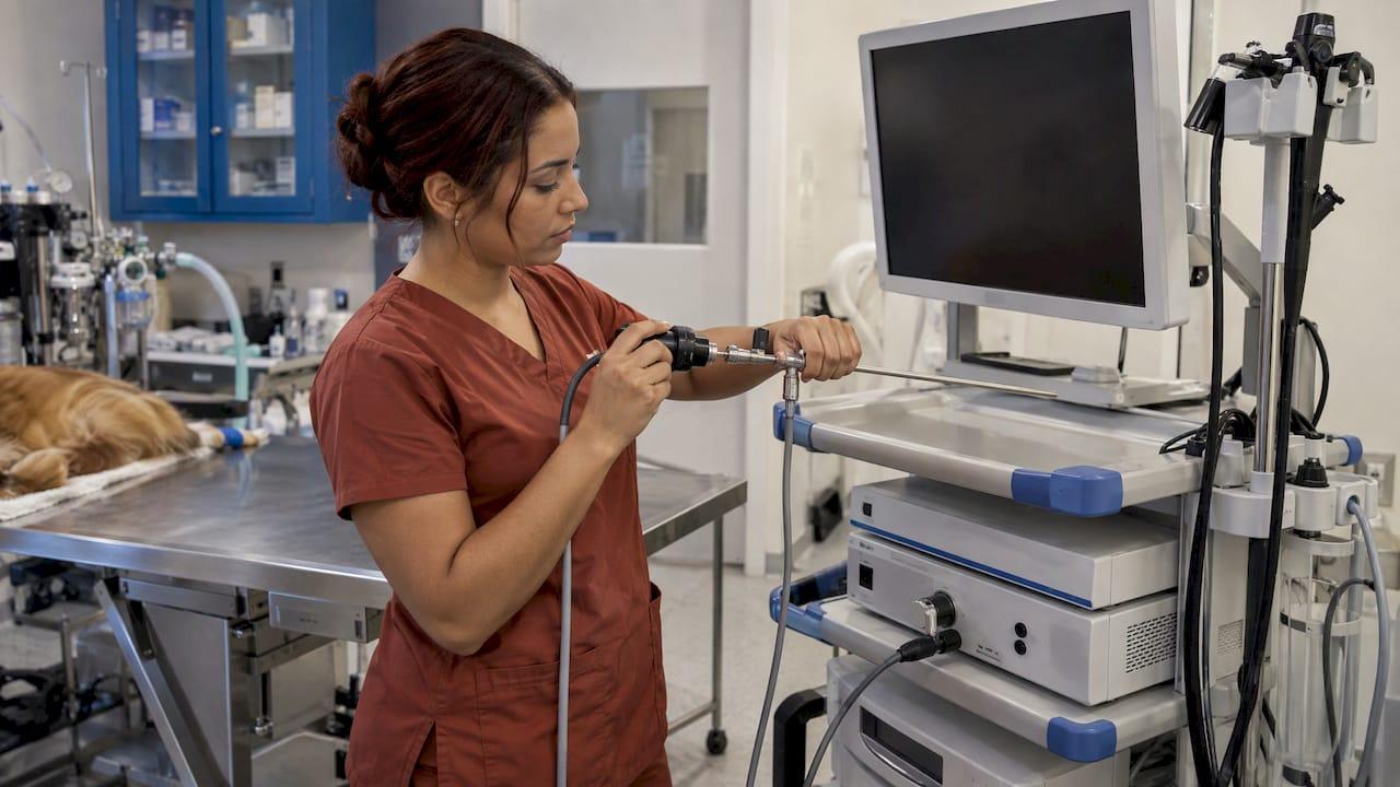
A veterinary urethroscopy setup has five parts: a scope sized to the patient, a light source, a camera and monitor, fluid management, and instruments for biopsy or retrieval. Add a reprocessing routine that fits the scope. The scope choice drives everything else. Female dogs and cats are generally examined with rigid scopes. Male dogs need a slim flexible scope. This guide covers sizes, components, accessories and cleaning so you can match equipment to your caseload.
TL;DR
- Urethroscopy looks at the urethra and is usually done together with cystoscopy of the bladder. Sizing is by patient: rigid scopes for females, small-diameter flexible scopes for male dogs (VCA Dallas).
- Common rigid sizes run from 1.9 mm for cats to 4.0 mm for medium-to-large female dogs. Male dogs generally need flexible scopes of about 2.5 to 3 mm.
- A complete system needs a light source, camera, monitor, fluid infusion and removal ports, and a working channel for biopsy forceps or retrieval tools.
- Flexible scopes are heat-sensitive. Clean and at least high-level disinfect them after every patient, following the maker's instructions.
- Keep more than one scope size on hand. A scope that is too large for the patient is a common reason the urethra cannot be entered.
Table of Contents
- What urethroscopy is, and how it relates to cystoscopy
- Rigid or flexible: choosing the scope type
- The components of a complete setup
- Matching equipment to your caseload
- Our perspective
- Cleaning, disinfection and storage
- A starter checklist for a urethroscopy cart
- Where to find equipment
- FAQ
- Sources
What urethroscopy is, and how it relates to cystoscopy
Urethroscopy is the endoscopic examination of the urethra. Cystoscopy examines the lining of the bladder. In practice the two are usually performed in one session, which is why equipment is often sold and discussed as cystoscopes, urethrocystoscopes or uroendoscopy systems. The same scope that views the bladder has to travel the length of the urethra first.
The approach differs by anatomy. VCA Dallas Animal Specialty notes that rigid scopes are generally used in female patients, and small-diameter flexible scopes in male dogs. Ureteroscopy, which goes further up into the ureter, is a separate and finer-scale procedure, so its scopes are narrower still.
The clinical reasons clinics reach for the scope are well established. VCA lists persistent hematuria, straining to urinate, vaginal discharge, removal of bladder or urethral stones, biopsy of a known bladder growth, urinary incontinence, and congenital urinary tract problems. DVM360 describes uroendoscopy as suited to lower urinary tract disease with unusual, prolonged or refractory signs, where it allows direct examination of the vestibule, vagina, urethra and bladder.
Those indications set your equipment requirements. If you only want to look, you need good optics and light. If you want to biopsy a mass or retrieve a stone, you need a working channel and the right instruments through it.

Rigid or flexible: choosing the scope type
Rigid scopes for female dogs and cats
A rigid cystoscope has three parts: a telescope, a sheath and a bridge. DVM360 explains that the glass fiber telescope can give an angled view of 0°, 12°, 30° or 70° from the tip, and the authors prefer 30°. The sheath carries irrigation and operating channels, and the bridge connects the light source and camera.
BSAVA illustrates two common configurations: a 4.0 mm × 30 cm telescope with a separate sheath and bridge, and a 2.7 mm × 18 cm telescope with an integrated sheath and bridge. The integrated design is simpler to assemble. The separate design gives more flexibility in how the sheath is used.
For cats, Berent describes a commonly used 1.9 mm 30° endoscope with a 3 Fr working channel for female cat access, whether vaginal, urethral, bladder or ureteral.
Flexible scopes for male dogs
The male dog urethra is long and curves around the os penis, so a slim flexible scope is the practical answer. DVM360 gives an example of a flexible endoscope with a 2.5 mm outer diameter (7.5 Fr), a 70 cm working length and a 1.2 mm (3.6 Fr) biopsy channel.
A clinical reference on dog urethra and bladder endoscopy lists the advantages of flexible instruments: fewer traumatic incidents and the ability to bend the tip to see the whole bladder interior. The same reference recommends flexible scopes up to 3 mm for males and for females of small and medium breeds, and up to 5 mm for females of large breeds.

Male cats are a special case. Berent notes that rigid cystourethroscopy from an antegrade approach is possible in male cats but does not allow imaging of the penile urethra.
Sizing at a glance
| Patient | Scope type | Example size | Source |
|---|---|---|---|
| Medium-to-large female dog | Rigid | 4.0 mm × 30 cm | DVM360 |
| Small-to-medium female dog | Rigid | 2.7 mm × 18 cm | DVM360 |
| Female cat, or male cat with perineal urethrostomy | Rigid | 1.9 mm × 18 cm | DVM360 |
| Male dog | Flexible | About 2.5 mm OD, 70 cm length | DVM360 |
These are examples from clinical references, not a rule for every patient. Individual anatomy varies, and a reference to cystoscopy techniques points out that one reason for failing to enter the urethra is a scope too large for the patient, which is why many practices keep several rigid sizes.
The components of a complete setup
Light source, camera and monitor
DVM360 states that rigid systems require a light source, a camera and a video monitor, preferably with image capture, and recommends xenon light with automatic intensity adjustment. Image capture matters more than it seems. A saved image or clip documents the lesion for the record, helps you explain findings to the owner, and supports referral or follow-up comparisons.
For flexible scopes, the light and camera are often integrated into the instrument or its video processor. Portable videoscope systems with a built-in monitor and SD card recording are practical when the same unit has to move between a procedure room and a barn or mobile van.
Fluid management
Urethroscopy is done with a distending fluid, so the scope needs ports to match. DVM360 says the scope needs ports for fluid infusion and removal and an instrument port. For a full urethral examination, the cystoscope must also allow retrieval of uroliths and foreign bodies and biopsy of masses.
Berent describes the rigid cystoscope as having three ports: a working channel plus ingress and egress ports. Fluids attach to ingress and an extension set to egress. She also notes the need for a good-quality cap on the working channel so fluid does not run out when the channel is open for an intervention.
Two practical points from the same article:
- Narrow channels restrict flow. Berent prefers a pressure bag with warm sterile saline (1 L) to keep fluid moving.
- Do not overdistend the bladder. She cautions that it could cause excessive bleeding and potentially bladder rupture.
One caution applies to flexible scopes. Their channels are small, and DVM360 warns against using mechanical suction because it could collapse the channel. Use flush and retrieval through the biopsy channel instead.
Working-channel instruments
The channel is only as useful as what passes through it. For a urethroscopy cart, plan on:
- Biopsy forceps sized to the channel. The 1.2 mm (3.6 Fr) channel in DVM360's flexible example, for instance, limits you to very fine instruments. VCA notes that biopsies can be passed alongside the scope, through its protective sleeve, or through an opening built into it.
- Retrieval baskets or graspers for stones and foreign material.
- Sterile saline, administration sets, a pressure bag and extension tubing.
- Spare channel caps and seals, which wear with use.
Our guide to biopsy forceps for small animal clinics covers 1.8 mm and 2.4 mm forceps and how they relate to channel size.
Matching equipment to your caseload
Start with who comes through the door, not with a catalog. A few questions narrow the list quickly.
- What is your mix of species and sexes? A practice seeing mostly female dogs and cats can build around rigid telescopes. If male dog urethral work is common, a slim flexible scope belongs on the list.
- What size range of patients? Toy breeds and cats sit at the 1.9 mm end. Large female dogs can take 4.0 mm. Many clinics end up with two or three rigid sizes.
- Diagnostic only, or therapeutic too? If you plan stone retrieval or biopsy, confirm the working channel diameter before you buy forceps or baskets.
- Shared or dedicated equipment? If a scope also handles other procedures, confirm the connectors and light guide are compatible.
- Where does the work happen? A fixed procedure room can use a cart. A mobile or multi-site practice benefits from a portable, monitor-equipped unit.
Anesthesia planning is part of the equipment conversation too. The dog urethra and bladder reference states that endoscopic examination of the urinary system is performed under general anesthesia, with dietary preparation of 24 hours of starvation and a 6-hour break from fluids. Your anesthesia protocol and monitoring belong to your clinical team, so confirm specifics with your anesthesiologist or internist.
For more on diameters across species, see our endoscope diameter guide and our overview of small diameter endoscopes in veterinary practice.
Our perspective
Most clinics we talk to do not need a hospital-grade human urology tower to start doing good urinary endoscopy. They need the right sizes, a reliable image, a working channel that fits their forceps, and a setup they can afford to maintain.
We built our business around veterinary-specific endoscopy at more affordable prices than traditional hospital-grade or human-use systems. That means portable, video-capable scopes with SD card recording, rigid scopes, flexible biopsy forceps and accessories such as valves, pumps and cleaning tools, all in one place, with 24/7 support and rapid shipping.
Our advice is to think in sizes and workflow before brands. Decide which patients you scope most, buy for that group first, and add sizes as your caseload grows. When a scope needs service, our repair service keeps equipment working instead of sitting in a drawer.
Endoscope
Cleaning, disinfection and storage
A scope that passes through the urethra and into the bladder is reprocessed every time. The CDC describes five steps after leak testing: clean, disinfect, rinse, dry and store. Cleaning includes brushing and flushing internal channels. High-level disinfectant is perfused through accessible channels for the product-recommended time. Heat-sensitive endoscopes must be cleaned and at least high-level disinfected after each use. The CDC also notes that contaminated endoscopes have been linked to outbreaks in healthcare settings.
A review of flexible-endoscope infection control describes the same multistep reprocessing: leak testing, cleaning, disinfection or sterilization according to manufacturer instructions, and storage that prevents recontamination.
For veterinary practices, the AAHA infection control guidelines add useful points:
- Cleaning removes visible organic matter with soap or detergent. Disinfection comes after and depends on that prior cleaning.
- Disinfectant contact times are generally 5 to 10 minutes, depending on the product concentration and target pathogens.
- Equipment that touches patients should be cleaned and disinfected between patients.
AAHA also notes that for endoscopy equipment that does not tolerate autoclaving, high-level disinfectants such as glutaraldehyde may be used. Always follow your scope maker's reprocessing instructions and the disinfectant label.
For a step-by-step routine, read our guides on endoscope sterilization for vets and sterilization in veterinary endoscopy. If your image dims or a channel behaves oddly, the videoscope troubleshooting guide is a good first stop.
A starter checklist for a urethroscopy cart
Use this as a working list when you plan a purchase or audit what you already own.
| Item | What to confirm |
|---|---|
| Rigid telescope(s) | Diameter and length for your smallest and largest female patients; 30° view is the angle the DVM360 authors prefer |
| Flexible scope | Outer diameter and working length for male dogs; biopsy channel size |
| Light source | Intensity control; compatible light guide |
| Camera and monitor | Image capture or SD recording |
| Fluid setup | Warm sterile saline, pressure bag, ingress and egress tubing, channel cap |
| Instruments | Forceps and retrieval tools matched to channel diameter |
| Reprocessing supplies | Leak tester, brushes, high-level disinfectant, rinse water, drying and storage |
| Service plan | Who repairs and calibrates the scope when it needs attention |
Where to find equipment
We supply veterinary endoscopy equipment and accessories for clinics, and you can start with our rigid scopes, arthroscopes and otoscopes. Our flexible biopsy forceps and endoscopic accessories round out a cart, and our repair service supports scopes already in your practice.
Tell us the species, sex and size range you scope, and our team can point you to the right category.
Browse the full range on our main catalog and place your order online.
FAQ
What is the difference between urethroscopy and cystoscopy?
Urethroscopy examines the urethra, and cystoscopy examines the bladder lining. They are usually performed together with the same scope, so the equipment is largely shared. Sizing is driven by how narrow the urethra is in the patient.
Can one scope handle both dogs and cats?
Usually not across the full range. References show different sizes for different patients: 4.0 mm for medium-to-large female dogs, 2.7 mm for small-to-medium female dogs and 1.9 mm for cats. Male dogs generally need a slim flexible scope. Practices that see both often keep several sizes.
Do I need a working channel for urethroscopy?
If you plan to biopsy or retrieve stones, yes. The scope needs ports for fluid infusion and removal and an instrument port for biopsy or retrieval tools. A diagnostic-only exam can be done with the viewing and irrigation functions alone.
How should a flexible urethroscope be cleaned after each patient?
Follow the CDC sequence after leak testing: clean, disinfect, rinse, dry and store. Brush and flush the channels, perfuse high-level disinfectant for the recommended contact time, and store the scope so it cannot be recontaminated. Your manufacturer's instructions take precedence.
Sources
- Cystoscopy, VCA Dallas Animal Specialty
- Rigid endoscopy: urethrocystoscopy and vaginoscopy, BSAVA
- Cystourethroscopy in the cat, Berent 2014
- Endoscopic examination of the urethra and the urinary bladder in dogs
- Uroendoscopy (Proceedings), DVM360
- Overview of small animal urinary tract endoscopy (Proceedings), DVM360
- Diagnostic approach to lower urinary tract disease (Proceedings), DVM360
- Cystoscopy: Techniques and clinical applications
- Disinfection of Healthcare Equipment, CDC
- Gastrointestinal Flexible Endoscopes: Infection Control
- 2018 AAHA Infection Control, Prevention, and Biosecurity Guidelines
- Sterilization methods in vet med, AAHA