
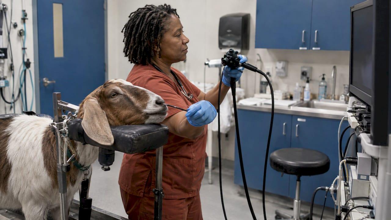
Endoscopy in sheep and goats works best when the scope fits the animal and the procedure. A flexible scope about 4 mm wide and 100 cm long covered the upper airway and esophagus in adult sheep and goats in a published study. Rigid telescopes handle vaginoscopy and laparoscopy. Sedation, fasting and regurgitation control matter as much as the optics. This guide covers each of those points for veterinarians, clinic managers and anyone buying equipment for a mixed or livestock practice. It also shows where our portable veterinary scopes, rigid scopes and biopsy forceps fit.
TL;DR
- Flexible endoscopy of the upper airway and esophagus is practical in adult sheep and goats. One study of 60 animals found the exam took about 10 minutes per animal with no major complications.
- Scope diameter is the first purchase decision. A 4 mm scope suited adults but was probably too large for animals under 8 months.
- Rigid telescopes with a working channel or port suit reproductive work such as vaginoscopy and targeted biopsy.
- Ruminants regurgitate under general anesthesia, so fasting, cuffed intubation and a clear sedation plan belong in every protocol.
- Portable video systems, rigid scopes, biopsy forceps and repairs are available from 1800endoscope for clinics that want one source for the whole setup.
Table of Contents
- What endoscopy covers in sheep and goats
- Upper airway and esophageal endoscopy
- Reproductive endoscopy: vaginoscopy and transcervical access
- Sedation, fasting and anesthesia
- Scope selection: size, length and type
- Biopsy and sampling
- A basic workflow for an airway exam
- What to look for when buying equipment
- Where 1800endoscope fits
- Our perspective
- FAQ
- Sources
What endoscopy covers in sheep and goats
Small ruminant endoscopy is the visual examination of internal structures through a scope. A camera and light are carried on a flexible or rigid tube. In sheep and goats, practice falls into four areas:
- Upper respiratory tract: nasal passages, pharynx, larynx, trachea and bronchi.
- Esophagus: often examined in the same session as the airway.
- Reproductive tract: vaginoscopy, transcervical work and laparoscopy.
- Abdominal laparoscopy: an alternative to exploratory laparotomy.
The core argument for the tool is simple. It lets you see the lesion rather than infer it from clinical signs. It also lets you take a targeted sample from it. Researchers who examined the upper airway and esophagus of 30 sheep and 30 goats concluded that the technique supports a more accurate diagnosis and prognosis. They recommended it as an adjunctive imaging modality.
This article is written for people who perform or equip these procedures. It does not cover owner-facing questions about what to expect for a pet.
Upper airway and esophageal endoscopy
The best-documented small ruminant procedure is flexible rhinolaryngotracheobronchoscopy plus esophagoscopy. In the study by Stierschneider, Franz and Baumgartner, the team examined 30 clinically normal sheep and 30 clinically normal goats aged 8 months to 8 years. They used a flexible endoscope 100 cm long and 4 mm in diameter. Key findings:
- The exam took about 10 minutes per animal.
- Sedation was needed in 8 of the sheep and 3 of the goats. Most animals tolerated the exam without it.
- No major complications were encountered.
- The distal lens sometimes steamed up and blurred tracheal images.
That last point is practical. Fogging is a handling issue you can plan for with a warm lens and a clean, dry insertion path. If your video system gives you trouble at the carina, our troubleshooting guide for animal endoscopy covers the common causes of poor images.
Which diseases justify the exam
The Merck Veterinary Manual lists sinusitis, nasal foreign bodies, nasal tumors and pharyngeal and laryngeal disorders among the upper respiratory diseases of sheep and goats. It also describes laryngeal chondritis, an obstructive upper-airway disease. It is most common in meat-breed rams and bucks 18 to 24 months old. It causes severe dyspnea from arytenoid cartilage edema. The manual states that endoscopic evaluation can diagnose it, while radiography and ultrasonography may show abscesses or calcification of the affected cartilages.
The practical rule: when a small ruminant has noisy breathing, exercise intolerance, nasal discharge or suspected obstruction, direct visualization often settles what imaging only suggests. The same group of authors noted that earlier reports have used the approach for nasal adenocarcinoma, adenopapilloma and laryngeal chondritis in sheep.
Reproductive endoscopy: vaginoscopy and transcervical access
Reproductive work is where rigid optics earn their place.
Transcervical cannulation in goats. A 2014 study of 15 Nigerian dwarf and African pygmy goats compared blind cannulation with endoscopic guidance. With a rigid 4 mm stainless steel cannula and an external light, 4 of 7 euthanized females showed complications including cervical penetration and damage. The team then used a custom 2 mm, 24.3 cm, 0-degree endoscope with a 2.4 mm dilator and LED light for the remaining 5 procedures. There were no complications, because the uterus could be seen after the scope passed through the cervix.
The lesson for equipment is clear. In small does, scope diameter and length are set by the anatomy, so choose the smallest rigid optic that gives you a usable image.
Vaginoscopy in ewes. A 2017 Colorado State University study performed serial vaginoscopy in 8 healthy anesthetized ewes at days 1, 14 and 28. The team used a laparoscopic single-incision port with a 10 mm, 30-degree, 33 cm rigid telescope and CO2 insufflation at about 6 mmHg. The technique was well tolerated. It allowed complete vaginal examination, identification of the cervix and targeted biopsies. The ewes were monitored daily for 28 days and no adverse events occurred.
The authors added that excellent viewing could outweigh the equipment cost, especially in hospitals already set up for laparoscopy. If your practice already owns rigid telescopes, extending them to small ruminant reproductive work is a modest step.

Sedation, fasting and anesthesia
This is where protocols succeed or fail, and the evidence differs by procedure.
Airway and esophageal exams
Most animals in the airway study needed no sedation. Where it was needed, it was needed in a minority: 8 of 30 sheep and 3 of 30 goats. Plan for restraint that keeps the head and neck in line, and have sedation drawn up for the animals that resist.
Procedures under general anesthesia
Ruminants often regurgitate under general anesthesia, especially in Trendelenburg position, where the head is lowered. The CSU protocol for ewe vaginoscopy was built around that risk. It included:
- 24-hour food withholding
- phenylbutazone (10 mg/kg PO) and procaine penicillin (30,000 IU/kg SC) 30 to 60 minutes before the procedure
- induction with IV ketamine (3.3 mg/kg) and diazepam (0.1 mg/kg)
- cuffed tracheal intubation
- isoflurane (1.5 to 3%) in oxygen at 1.5 L/min
- lactated Ringer's at 3 to 5 mL/kg/h
These are the study's published doses for that procedure in healthy ewes. They are a reference point, not a universal protocol. The attending veterinarian sets drugs and doses for the individual animal, taking into account health status, species, local regulations and withdrawal times in food animals. The authors suggested that sedation-based restraint protocols warrant further study.
Laparoscopy
A 2020 chapter on small ruminant gastrointestinal disease notes that laparoscopy can serve diagnostically as an alternative to exploratory laparotomy. General anesthesia is recommended for better abdominal inflation, though sedation with local anesthesia at the portal sites is possible. Fasting for 24 to 48 hours improves visualization. Assisted ventilation should be available, because abdominal inflation and head-down positioning press on the diaphragm.
Scope selection: size, length and type
Before you compare video systems, settle three questions.
1. How big is the animal? The 4 mm, 100 cm flexible scope worked in adult sheep and goats but was probably too large for animals under 8 months. If you see lambs and kids, you need a smaller diameter. Our overview of small diameter endoscopes and their veterinary uses explains the trade-offs between diameter, image and working length.
2. Flexible or rigid? Match the optic to the procedure:
| Procedure | Scope type used in the research | Diameter in the study |
|---|---|---|
| Airway and esophagus, adult sheep and goats | Flexible, 100 cm | 4 mm |
| Transcervical access, small goats | Rigid, 0 degree, 24.3 cm | 2 mm |
| Vaginoscopy, adult ewes | Rigid, 30 degree, 33 cm, with laparoscopic port | 10 mm |
3. Where will you use it? Small ruminant work often happens in a barn, a pen or at a client's farm. A portable system with a direct monitor and SD card recording lets you capture the exam without a cart or a wall outlet setup. Video also helps with second opinions and client records, and our guide to optimizing endoscope video recording covers settings that matter most.
Biopsy and sampling
Visualization tells you where to sample. Endoscopic biopsy turns a visual finding into a histopathology result. In the ewe vaginoscopy study, the technique allowed targeted biopsies without complications. The same logic applies to nasal masses and pharyngeal lesions, where a guided forceps sample beats a blind one.
Two habits improve the result:
- Take several samples from the lesion and from adjacent tissue, not a single bite.
- Confirm that the forceps fits the working channel of your scope before the day of the procedure.
Our flexible biopsy forceps page lists the options. Our guide to GI endoscope specifications explains how channel size and optics affect sample quality, and it helps you avoid repeat biopsies.
A basic workflow for an airway exam
This sequence reflects what the published study did, with practical points added.
- Check the equipment. Confirm light output, image, insertion tube condition and that the scope passes cleanly through your lubrication and cleaning routine.
- Restrain. Keep the head level and steady. Have sedation ready.
- Enter through the nostril. Examine the nasal passages, then the pharynx and larynx.
- Assess the larynx. Look at arytenoid cartilage symmetry and swelling, the finding that matters in laryngeal chondritis.
- Advance to the trachea and bronchi. Expect some lens fogging and wipe or warm the tip as needed.
- Examine the esophagus on withdrawal or in a second pass.
- Record the exam. Save images and video to the patient file.
- Clean and reprocess the scope before the next animal.
The study reported about 10 minutes per animal, so the whole sequence is realistic to include in a farm call.
What to look for when buying equipment
Procurement for a livestock or mixed practice comes down to six points:
- Insertion tube diameter that matches your smallest patients.
- Working length long enough to reach the carina or distal esophagus in an adult.
- Portability and power, so you can work in a barn.
- Video capture to SD card or PC.
- Accessory compatibility, including biopsy forceps, valves, pumps and light sources.
- Repair access. Insertion tubes take a beating around animals, and a repair route keeps a scope in service.
Our rigid scopes, arthroscopes and otoscopes cover the reproductive and ear work. Our airway field scopes include the portable 9.5 x 150 airway inspection videoscope and the 8 mm equine airway field scope, and the portable 12.9 x 3.5 meter gastroscope is for longer reach. Our equine guide to airway endoscopy equipment explains how the same airway principles apply across species.
Where 1800endoscope fits
We sell veterinary endoscopes, videoscopes, light sources, biopsy forceps and accessories such as pumps, valves and cleaning tools. We also repair endoscopes. Our systems are portable, and several records to an SD card, so a practice can build a small ruminant setup from one catalog, with support available 24/7 and quick shipping.
If you are equipping a practice for sheep, goats and other livestock, start with the veterinary endoscopy equipment page. For the full range, browse the 1800endoscope catalog.
When you want the scope, forceps and accessories matched to one another, the medical endoscopes and accessories page is the place to place an order. It includes our New Portascope.
Our perspective
Small ruminant work rewards practices that keep the equipment simple. The published studies point the same way: a modest flexible scope for the airway and esophagus, a small rigid optic for reproductive access and a reliable way to record what you see.
We build our catalog for that kind of practice. Veterinary clinics should not have to pay for hospital-grade human systems to examine a ewe or a doe. Portable videoscopes, rigid scopes, biopsy forceps and the accessories around them let a clinic go from the clinic to the barn with one kit, and repairs keep that kit working.
The studies also show that technique matters as much as the tool. Fasting, sedation planning and correct scope size decide whether an exam is quick and uneventful. Pair the right equipment with a protocol your team has rehearsed, and endoscopy becomes a routine part of a livestock workup rather than a special event.
Endoscope
FAQ
Can you perform endoscopy on sheep and goats without general anesthesia?
For upper airway and esophageal exams in adults, often yes. In the 60-animal study, sedation was needed in only 8 sheep and 3 goats. Procedures that need abdominal or pelvic inflation, such as laparoscopy and ewe vaginoscopy, are described with general anesthesia, though laparoscopy with sedation and local anesthetic is possible.
What size scope do you need for a sheep or goat?
A flexible scope 4 mm wide and 100 cm long worked in adults aged 8 months and older, and was probably too large for younger animals. For transcervical work in small goats, researchers used a 2 mm rigid endoscope. Measure your smallest planned patient before choosing.
Which conditions can endoscopy help diagnose in small ruminants?
Upper airway problems are the clearest use: sinusitis, nasal foreign bodies, nasal tumors and pharyngeal or laryngeal disorders. Laryngeal chondritis in rams and bucks can be diagnosed endoscopically. Endoscopy can also help confirm and localize pharyngeal trauma and abscesses, alongside radiographs and ultrasound.
Why do protocols stress fasting and cuffed intubation?
Ruminants often regurgitate under general anesthesia, particularly with the head lowered. Fasting for 24 hours or more and cuffed tracheal intubation reduce the risk of aspiration. Your veterinarian sets the exact plan for each animal.
Sources
- Endoscopic examination of the upper respiratory tract and esophagus in sheep and goats
- Overview of Respiratory Diseases of Sheep and Goats, Merck Veterinary Manual
- Successful Use of Endoscopy for Transcervical Cannulation in goats
- Vaginoscopy in Ewes Utilizing a Laparoscopic Surgical Port
- Diseases of the gastrointestinal system, small ruminant chapter