
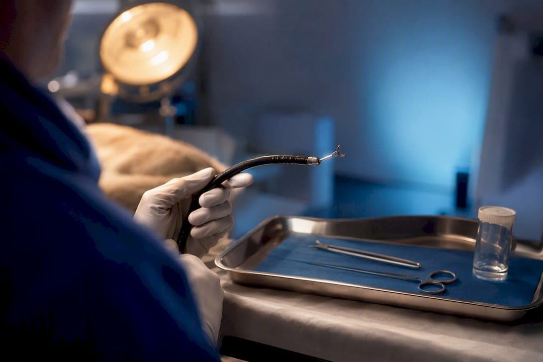
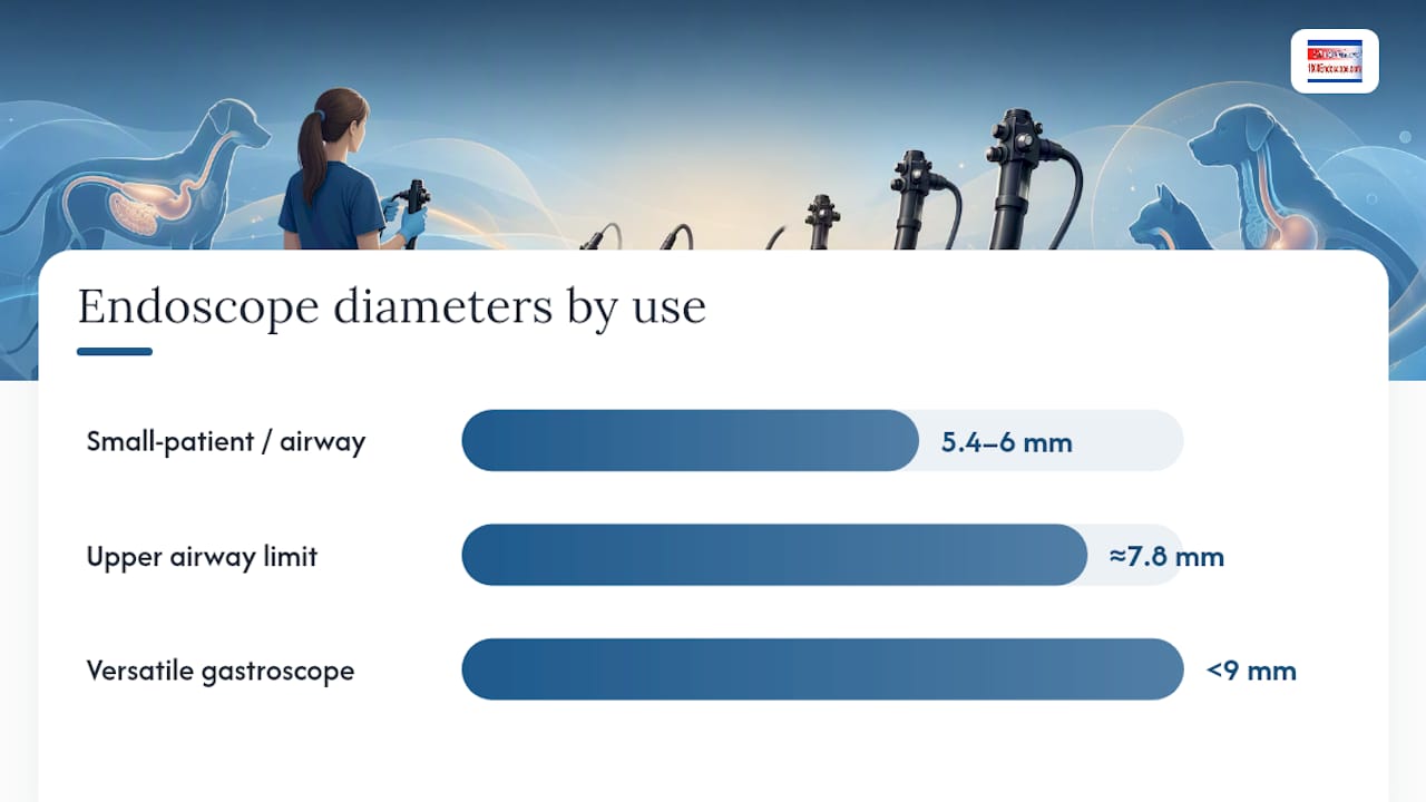
For most canine and feline GI cases, the right tool is a flexible videoendoscope of suitable diameter and length, typically equipped with a biopsy channel large enough to accept standard biopsy forceps. That single instrument covers esophagoscopy, gastroscopy, and duodenoscopy in the vast majority of patients you'll see. Cats, toy-breed dogs, and any clinic doing airway work alongside GI cases need a second, slimmer scope in the 5.4 to 6 mm range. Portable systems with small-diameter units now cover a meaningful share of that need, without a cart-based system's price tag or footprint.
TL;DR:
- Smaller-diameter scopes in the 5.4 to 6 mm range are essential for airway and urethral procedures alongside GI work in small animals, requiring a second scope budget.
- A biopsy channel of at least 2.8 mm is necessary to obtain sufficient tissue samples for reliable histopathology, reducing the need for repeat procedures.
- Proper scope reprocessing, including thorough drying and inspection after each use, is critical to prevent fogging, biofouling, and costly equipment damage.
- A portable endoscopy system like the PVSM6150 provides effective diagnostic upper GI work in small practice settings without the expense or footprint of a cart-based unit.
- Videoendoscopes offer superior image quality, easier documentation, and more reliable long-term performance than fiber scopes, influencing initial purchase choices.
Table of Contents
- What GI Endoscopy Actually Diagnoses and Treats in Small Animals
- Flexible vs. Rigid Endoscopes: Matching the Scope to the Job
- Planning the Procedure: Prep, Sedation, and Biopsy Handling
- Fixing Fogging and Biofouling Mid-Procedure
- Running Endoscopy Day Smoothly: Team and Documentation
- How 1800endoscope Thinks About Small-Animal GI Equipment
- A Portable Option Worth Considering for GI Work
- Sources
What GI Endoscopy Actually Diagnoses and Treats in Small Animals
Endoscopy is a minimally invasive way to look inside the GI tract and, in many cases, act on what you find without opening the abdomen. It works because flexible scopes are built for tubular anatomy, threading through the esophagus, stomach, small intestine, and colon in a way that rigid instruments simply cannot, according to PetMD's overview of veterinary endoscopy. Rigid endoscopes have their place in the practice too, mainly for joints, the abdominal cavity, nasal passages, and ears, but the GI tract belongs to flexible scopes.
The three procedures that make up most small animal GI endoscopy caseloads each answer a different clinical question:
- Esophagoscopy evaluates strictures, esophagitis, vascular ring anomalies, and foreign bodies lodged in the esophagus, and it doubles as a retrieval tool for many of those foreign bodies.
- Gastroscopy and duodenoscopy assess chronic vomiting, gastric ulceration, foreign bodies, and infiltrative disease like lymphoma or inflammatory bowel disease, and they let you collect mucosal biopsies without surgery.
- Colonoscopy and ileoscopy work up chronic large-bowel diarrhea, colitis, polyps, and neoplasia, and they reach the ileum in many but not all patients depending on scope length and patient size.
Vomiting, diarrhea, unexplained weight loss, suspected foreign body ingestion, and chronic mucosal disease are the indications that land most cases on your schedule. A straightforward gastroscopy for a vomiting adult dog is routine outpatient work in general practice. Complex therapeutic removals, sharp or linear foreign bodies deep in the small intestine, and advanced interventional procedures tend to move up to referral centers with more scope inventory and anesthesia support. University hospital services, like the one at UF's Small Animal Hospital, routinely list upper and lower GI endoscopy alongside interventional foreign body retrieval as standard offerings, which gives you a useful benchmark for what "referral level" actually looks like.
Recovery logistics matter for how you counsel owners. Diagnostic endoscopy is typically a same-day procedure. Patients go home once they're stable after sedation, according to South Sacramento Pet Hospital's overview of dog and cat endoscopies. Biopsy results, on the other hand, take days to come back from the lab, so the procedure itself resolves fast but the diagnosis often doesn't.
Flexible vs. Rigid Endoscopes: Matching the Scope to the Job
Flexible fiberscopes and videoendoscopes dominate small animal digestive endoscopy for one simple reason: the GI tract bends, and your instrument has to bend with it. Fiberscopes use bundled glass fibers to transmit an image to an eyepiece or camera adapter. Videoendoscopes put a chip at the tip and send a digital signal straight to a monitor. Rigid telescopes, by contrast, are straight, non-deflecting instruments best suited to non-tubular spaces like joints or the abdominal cavity, not the twists of a stomach or colon.
Here's how the two flexible categories stack up against each other:
- Image quality. Video chips generally deliver sharper, more consistent images than fiber bundles, especially as fiber optics age and individual fibers break, creating dark spots across the image.
- Documentation. Video systems record directly to SD card or hard drive for the medical record. Fiber systems need an external camera adapter to capture anything beyond a live look.
- Repair cost. Fiber scopes are generally cheaper to repair when a few fibers break. A damaged video chip is often a full head-assembly replacement.
- Longevity under rough handling. Fiber bundles tolerate some abuse before image quality degrades noticeably. Video chips fail more abruptly when they're damaged.
The specifics that actually govern purchase decisions come down to diameter, length, and channel size. Today's Veterinary Practice recommends an insertion tube under 9 mm and a working length of 100 to 125 cm as the most versatile all-around instrument for a general small animal practice. That length reaches the stomach and duodenum comfortably in cats and dogs up to roughly 40 to 50 pounds. Large-breed colonoscopy sometimes needs more than 125 cm to reach the proximal colon and ileum without excessive looping.
Biopsy channel size deserves its own line item on your spec sheet. A channel of at least 2.8 mm accepts standard biopsy forceps that pull adequate tissue samples for histopathology. Go smaller and you're stuck with cup forceps that grab too little tissue to get a reliable diagnosis, which means repeat procedures and unhappy owners.
Diameter isn't just about fitting through a small esophagus. A larger insertion tube carries more optical fibers or a bigger chip, which generally means better light transmission and a wider field of view. That's a real trade-off against trauma risk in a five-pound cat or a toy breed. Practices that regularly work on very small patients, or that also perform rhinoscopy, bronchoscopy, or urethrocystoscopy, need a second scope in the 5.4 to 6 mm range because anything larger than about 7.8 mm becomes unworkable for those airway and urethral procedures. 1800endoscope's small-diameter endoscope lineup covers exactly that gap for clinics that see a lot of cats and small dogs.
Pro Tip: Don't buy a single scope and try to make it do everything. A 9 mm gastroscope forced into a cat's trachea for a bronchoscopy is a good way to cause airway trauma and a bad day for everyone involved. Budget for the second, slimmer scope from the start if airway work is part of your caseload, and use a size selection guide to confirm fit before you order.
Planning the Procedure: Prep, Sedation, and Biopsy Handling
Reach varies more than most new endoscopists expect. A 110 cm scope in a 60 pound Labrador might not consistently reach the ileum, while the same scope in a 10 pound cat reaches well past the ileocecal junction with room to spare. Know your scope's real-world reach in your typical patient population before you promise an owner a full ileoscopy.
Prep protocols differ by procedure type:
- Upper GI (esophagoscopy, gastroscopy, duodenoscopy) needs a 12 hour fast to clear the stomach of food that would otherwise obscure the mucosa and clog the working channel.
- Lower GI (colonoscopy, ileoscopy) needs actual bowel prep, typically an oral lavage solution or repeated warm water enemas the evening before and morning of the procedure, since retained fecal material makes colonoscopy essentially useless.
- Anesthesia is general anesthesia for nearly all small animal GI endoscopy, not sedation alone. Patients need to hold still for scope passage and biopsy collection, and gagging or swallowing reflexes make sedation-only approaches impractical for anything beyond a quick look.
Biopsy technique is where a lot of diagnostic value gets won or lost. Take multiple samples, generally six to eight from a suspicious gastric or colonic lesion, since a single biopsy risks missing patchy disease like early lymphoma. Use forceps sized to your channel (that 2.8 mm minimum again) and orient samples on a piece of lens paper or a biopsy sponge before dropping them in formalin, so the pathologist gets a properly oriented section rather than a crushed, unreadable fragment. Label each container immediately with location and patient ID. It sounds obvious until you're juggling six samples from three different colon segments and a phone ringing in the next room.
Referral triggers are worth setting as a practice policy rather than deciding case by case. Deep small intestinal foreign bodies, suspected strictures needing balloon dilation, and any therapeutic intervention beyond straightforward biopsy or simple foreign body retrieval are reasonable lines to draw. Interventional capability, the kind UF's endoscopy service lists among its routine offerings, generally requires equipment and repetition that a general practice doing occasional endoscopy won't have on hand.
Fixing Fogging and Biofouling Mid-Procedure
Fogging happens when warm, humid patient tissue meets a cooler lens, and biofouling happens when blood, mucus, or debris coats the lens surface during the procedure. Both problems have the same practical effect: you lose visibility exactly when you need it most, often mid-biopsy or while chasing a foreign body around a gastric fold.
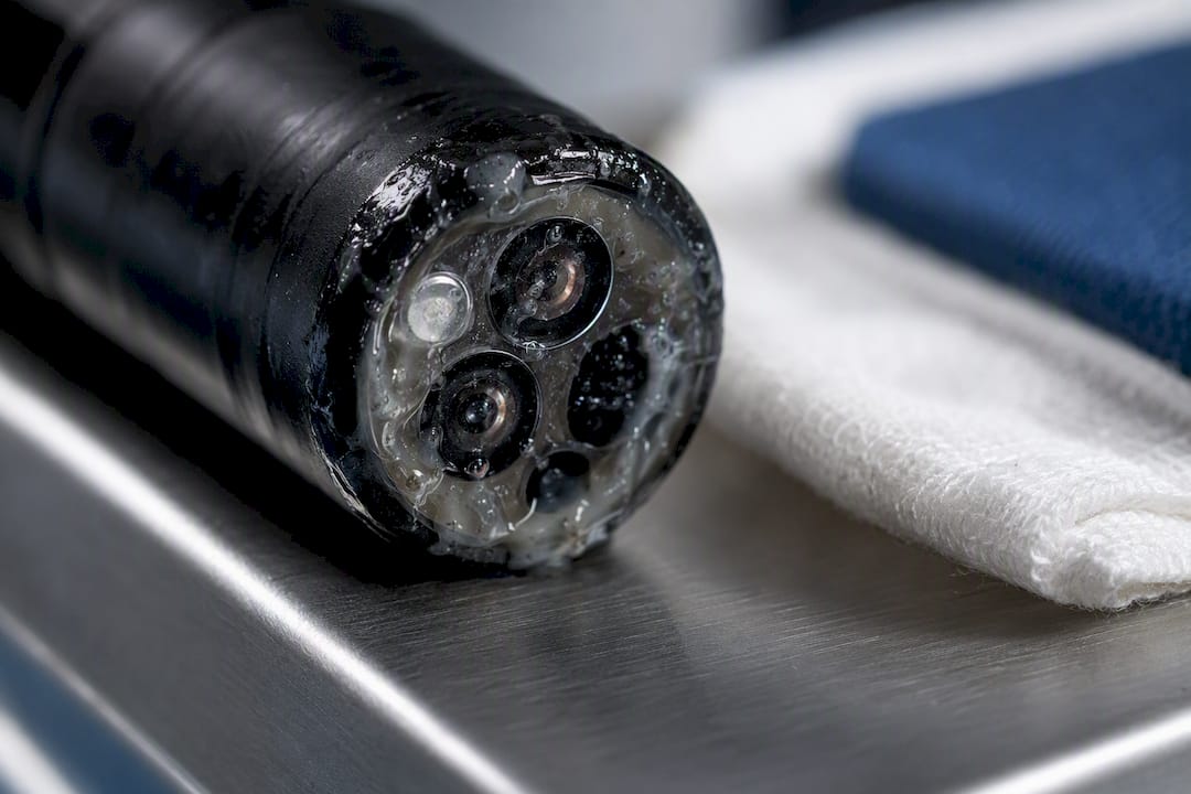
A controlled trial testing anti-fog options in head-and-neck endoscopy found that ordinary baby shampoo performed significantly better than using no agent at all, and held up comparably to commercial anti-fog products, according to research published in PMC. That's a genuinely useful finding for a small animal practice watching supply costs. A thin film of diluted baby shampoo applied to the lens tip before insertion is a low-cost, evidence-backed step, not a folk remedy.
During the procedure itself, a few habits keep you ahead of the problem:
- Use your irrigation and suction channels proactively rather than waiting until the image is already obscured.
- Withdraw and wipe the lens the moment image quality starts to degrade rather than pushing through a hazy view and risking a missed lesion or a biopsy taken from the wrong spot.
- Clean the lens tip gently with a soft, non-abrasive swab. Scrubbing at debris with anything stiff risks scratching the lens coating and creating a permanent visibility problem.
Emerging lens technology is worth watching but not worth waiting for. Lubricant-infused engraved nano-microstructures, described in research on durable anti-fogging endoscope lenses published in Scientific Reports, showed strong anti-fogging and anti-biofouling performance in lab testing, including high visible-area retention through blood-spray and dipping simulations. That's promising in vitro data. It hasn't yet translated into widely available clinical scopes on veterinary equipment price lists, so budget and plan around today's baby-shampoo-and-good-technique reality rather than tomorrow's coating.
Running Endoscopy Day Smoothly: Team and Documentation
A GI endoscopy case runs better with defined roles rather than three people improvising around one table. Assign a scope operator focused entirely on navigation and biopsy collection, a table assistant handling instrument passing and sample labeling, an anesthesia tech monitoring vitals throughout, and a reprocessing lead responsible for cleaning and drying the scope immediately after the case ends.
Get your team comfortable with the equipment before it's an actual patient on the table:
- Start with in-house mentorship. Have your most experienced endoscopist run a junior tech or new associate through dry passes on a training model or a cadaver before their first live case.
- Add structured continuing education. Veterinary conferences and specialty societies run hands-on endoscopy wet labs that build real skill faster than reading alone.
- Document every case on video. Save clips for the medical record, for client communication when explaining findings, and for billing justification when insurance or owners question the procedure. 1800endoscope's technician tips resource covers workflow details worth building into your own checklist.
- Track scope condition after every use. Watch for degrading image quality, new dark spots (a sign of broken fiber bundles), or difficulty with tip deflection, since these are the red flags that mean repair time before the next case, not after.
The most common and costly mistakes aren't clinical, they're logistical: rushing reprocessing, skipping the drying step, or sending a scope back into service with a channel that wasn't fully flushed. Inadequate biopsy sampling from rushing the procedure is the other repeat offender, and it costs you a callback and a second anesthesia event far more often than it saves you five minutes.
How 1800endoscope Thinks About Small-Animal GI Equipment
1800endoscope stocks portable small-diameter systems and full accessory catalogs built around the specs that actually matter in practice: sub-9 mm gastroscopes, slimmer airway-capable scopes, and the biopsy forceps and valves that make a scope clinically useful rather than just a camera on a stick. Browse the endoscopy resource blog for use-case detail, or reach out directly for spec sheets and compatibility questions before you commit to a system.
— Endoscope
A Portable Option Worth Considering for GI Work
If your caseload is mostly diagnostic upper GI work, cats, small dogs, and general practice volume rather than a referral center's therapeutic caseload, a full cart system is often more equipment than you need. The PVSM6150, a fully portable 6 mm by 150 cm system with direct SD card video recording, gives you an insertion tube slim enough for small patients, a working length that reaches the stomach and duodenum comfortably, and recorded documentation for the medical record, all without a cart-based unit's price or footprint.
It has real limits. Complex large-breed colonoscopy reaching the proximal colon, or interventional work needing a wider biopsy channel and multiple accessory ports, still calls for a full cart system with more channel options. For everyday upper GI diagnostics in general practice, a portable unit like this covers a large share of what actually walks through the door. Browse the broader borescope and endoscope catalog for accessory options, or contact 1800endoscope directly to confirm compatibility with your current light source and monitor setup before you order.
Sources
Video versus fiber is the first fork in the road, and it shapes almost everything downstream. A digital videoendoscope gives you a chip-based image displayed on a monitor, recorded to SD card or internal storage, and easy to share with a specialist for a second opinion. A fiberscope gives you a dimmer, grainier eyepiece view unless you add a camera head, and that add-on itself becomes another point of failure.
Run through this checklist before you commit to a system:
- Endoscopic Equipment for the Veterinary Practitioner (Today's Veterinary Practice)
- Veterinary endoscopy (PetMD)
- Alternative agents to prevent fogging in head and neck endoscopy (PMC)
- Lubricant-infused directly engraved nano-microstructures for mechanically durable endoscope lens (Nature Scientific Reports)
- Endoscopy — UF Small Animal Hospital
Reprocessing deserves more weight in the buying decision than most clinics give it. Flash sterilization and inadequate drying before storage let moisture sit inside the scope's channels and housing, and that trapped moisture becomes internal condensation that fogs the lens from the inside and, over time, corrodes the optics permanently. A drying cabinet or a documented air-drying protocol after each cleaning cycle is cheap insurance against a repair bill that dwarfs the scope's original price.
Pro Tip: Before you buy, ask the vendor directly what reprocessing method the scope was validated against. A scope rated only for cold sterilization that gets run through your autoclave "just this once" is how a five-figure instrument turns into a five-figure paperweight. If you want a side-by-side on where a compact system covers your needs versus a full cart, 1800endoscope's recording optimization guide walks through documentation settings that apply to either setup.