
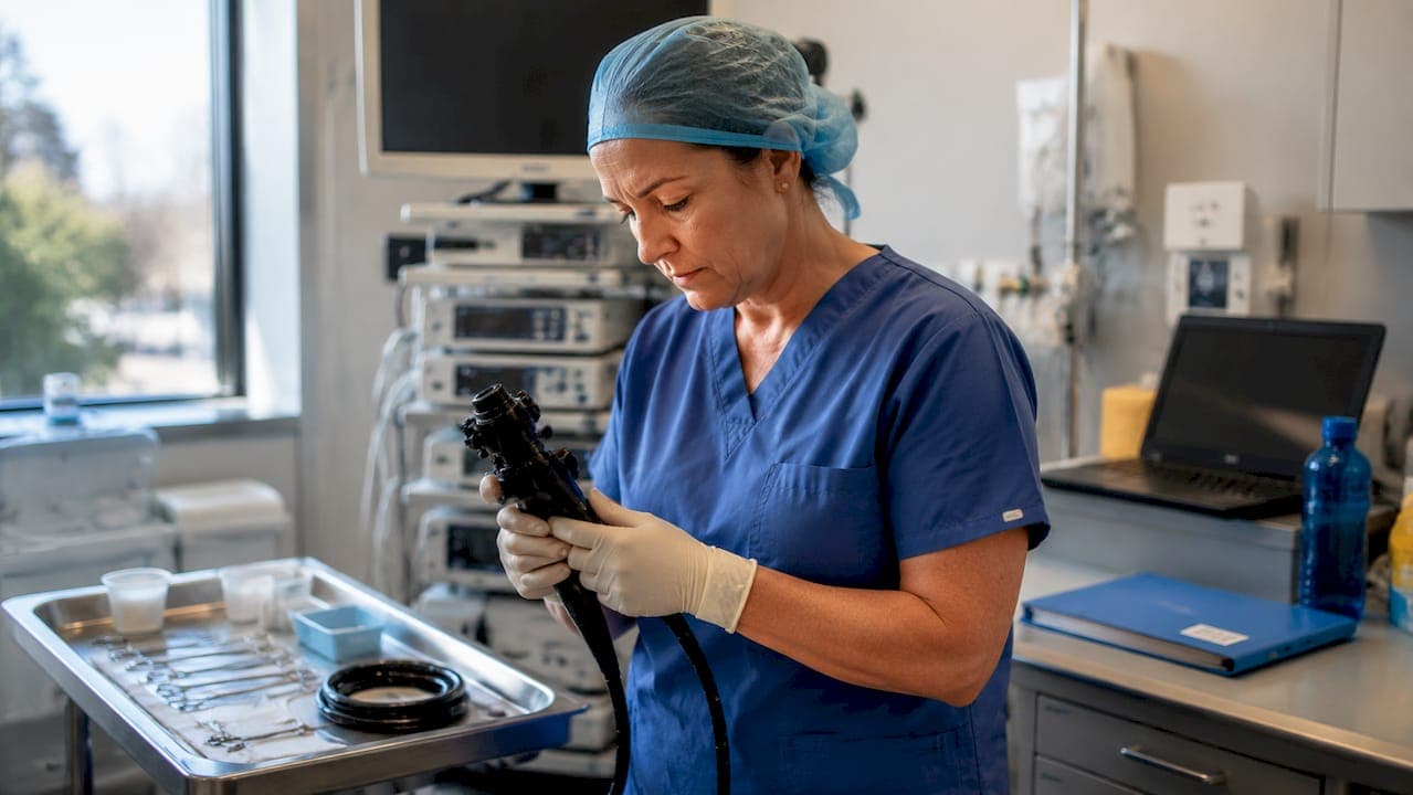
Common endoscopic procedures are minimally invasive techniques that use flexible or rigid instruments to visualize, diagnose, and treat conditions within body cavities. Endoscopy, the recognized clinical term, spans a broad range of applications from upper gastrointestinal tract examination to airway assessment and veterinary gastroscopy. The field has shifted decisively from purely diagnostic work toward therapeutic intervention, making procedural knowledge more critical than ever for both human medicine and veterinary practice. This guide covers the most clinically relevant types of endoscopy, their therapeutic applications, and the technology shaping modern practice.
What are the most common endoscopic procedures?
The endoscopic procedures list in human medicine is anchored by three high-volume techniques: esophagogastroduodenoscopy (EGD), colonoscopy, and endoscopic retrograde cholangiopancreatography (ERCP). Each targets a distinct anatomical region and serves both diagnostic and therapeutic roles.
Esophagogastroduodenoscopy (EGD) is the most frequently performed upper GI procedure. Approximately 6.1 million EGDs are performed annually in the United States. That volume reflects how central upper endoscopy is to diagnosing conditions like Barrett's esophagus, peptic ulcer disease, and gastric malignancy. A diagnostic EGD lasts roughly 15 minutes, though sedation preparation and recovery extend the total clinical encounter significantly.
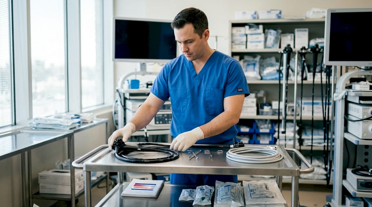
Colonoscopy is the standard tool for colorectal cancer screening. The American Cancer Society recommends colonoscopy starting at age 45 for average-risk adults. That recommendation reflects the procedure's dual power: it identifies suspicious lesions and removes them in the same session through polypectomy.
ERCP occupies a specialized tier. It combines endoscopy and fluoroscopy to access the biliary and pancreatic ducts, enabling stone extraction, stent placement, and sphincterotomy. ERCP demands advanced technical skill and carries a higher complication profile than standard gastrointestinal endoscopy.
Beyond these three, the common endoscopic exams list includes:
- Bronchoscopy: Direct airway visualization for biopsy, foreign body retrieval, and lavage
- Laryngoscopy: Assessment of vocal cord pathology, airway obstruction, and supraglottic lesions
- Cystoscopy: Bladder and ureteral examination for hematuria workup and tumor surveillance
- Thoracoscopy: Pleural space access for biopsy and effusion management
| Procedure | Primary Organ System | Typical Duration |
|---|---|---|
| EGD | Upper GI tract | ~15 minutes |
| Colonoscopy | Colon and rectum | 20–45 minutes |
| ERCP | Biliary/pancreatic ducts | 30–60 minutes |
| Bronchoscopy | Airways and lungs | 20–30 minutes |
| Cystoscopy | Bladder and urethra | 15–30 minutes |
How are endoscopic techniques used in veterinary practice?
Veterinary endoscopy mirrors human gastrointestinal endoscopy in principle but differs substantially in patient anatomy, sedation protocols, and equipment sizing. The most common veterinary endoscopic techniques include gastroscopy, bronchoscopy, rhinoscopy, and arthroscopy.
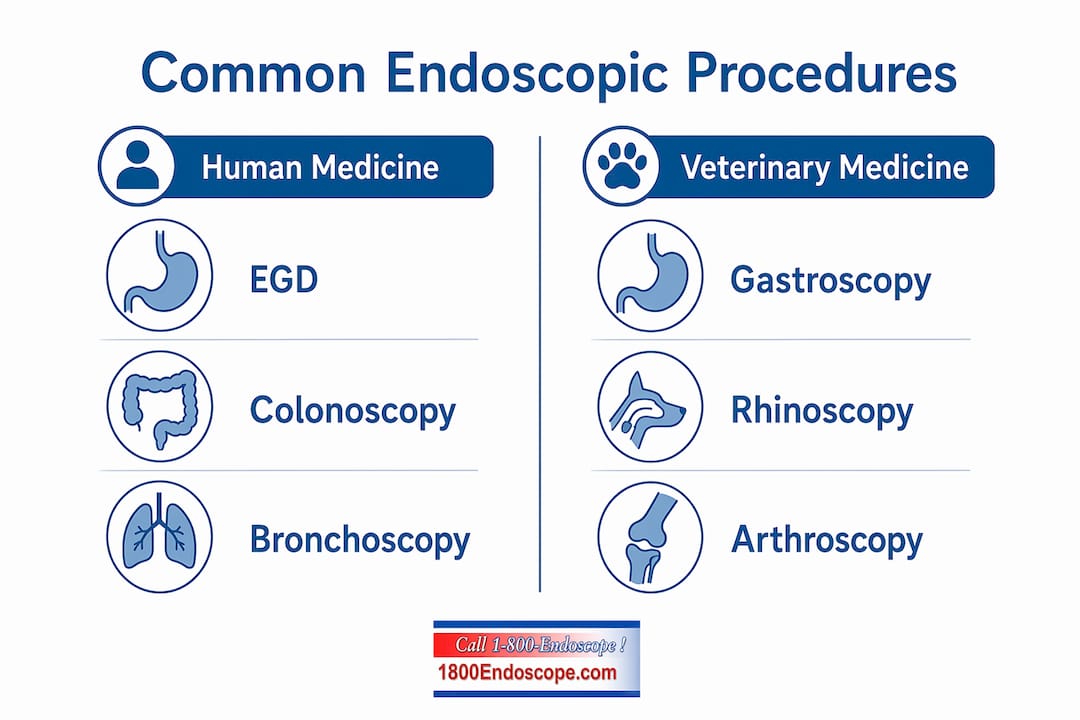
Gastroscopy in small animals uses flexible videoscopes to evaluate chronic vomiting, esophageal foreign bodies, and inflammatory bowel disease. In horses, gastroscopy is the definitive method for diagnosing equine gastric ulcer syndrome, a condition affecting a large proportion of performance horses. Equipment selection matters here. Small diameter endoscopes are preferred for cats and small dogs, where a 5–8 mm insertion tube diameter is standard.
Rhinoscopy allows direct visualization of the nasal cavity and nasopharynx in dogs and cats. It is the primary diagnostic tool for chronic nasal discharge, epistaxis, and suspected nasal tumors. Rigid rhinoscopes provide superior image quality for this application.
Bronchoscopy in veterinary patients follows the same anatomical logic as human bronchoscopy. It is used for bronchoalveolar lavage, foreign body retrieval, and assessment of tracheal collapse in brachycephalic breeds. Practitioners should review veterinary laryngoscopy protocols before approaching airway procedures in compromised patients.
Arthroscopy is the gold standard for diagnosing and treating joint pathology in dogs and horses. It provides magnified visualization of cartilage, synovium, and ligamentous structures that radiography cannot resolve.
Key advantages of minimally invasive endoscopic approaches in veterinary patients include:
- Reduced anesthesia time compared to open surgery
- Faster recovery and shorter hospitalization
- Ability to collect targeted biopsy samples without laparotomy
- Real-time video documentation for case records and client communication
Pro Tip: When performing veterinary gastroscopy, fast small animals for at least 12 hours and large animals for 24–36 hours before the procedure. Residual ingesta obscures mucosal detail and increases aspiration risk.
What therapeutic interventions do endoscopic procedures enable?
Endoscopy's therapeutic range has expanded well beyond biopsy collection. Polypectomy is the primary mechanism for colorectal cancer prevention, converting a screening colonoscopy into a cancer-preventing intervention in a single session. Removing adenomatous polyps before they progress to carcinoma is one of the most clinically impactful actions available in outpatient medicine.
Stricture dilation addresses narrowing in the esophagus, pylorus, or colon. Bougie dilators use mechanical force along a guidewire, while balloon dilators allow through-the-scope access with precise diameter control. Balloon dilation is preferred for most GI strictures because it applies radial force uniformly, reducing the risk of mucosal tearing.
Endoscopic stenting manages obstructions in the biliary tree, esophagus, and colon. Self-expanding metal stents are deployed under fluoroscopic or endoscopic guidance to restore luminal patency in malignant or benign obstructions. In palliative oncology, colonic stenting avoids emergency surgery and significantly improves quality of life.
Hemostasis is another critical therapeutic application. Endoscopic techniques for bleeding control include injection therapy with epinephrine, thermal coagulation using heater probes or argon plasma coagulation, and mechanical clip placement. Each method suits different bleeding sources and vessel sizes.
Foreign body removal is common in both human and veterinary endoscopy. In children and animals, ingested objects lodged in the esophagus or stomach are retrieved using rat-tooth forceps, snares, or retrieval nets passed through the working channel.
Endoscopy carries risks including perforation and significant bleeding, which require thorough informed consent and structured post-procedure monitoring. That risk profile is not a reason to avoid the procedure. It is a reason to prepare patients and staff rigorously.
Pro Tip: After any therapeutic endoscopic procedure involving dilation or polypectomy, monitor patients for at least 30 minutes for signs of perforation, including tachycardia, abdominal rigidity, and subcutaneous emphysema.
How does endoscopic technology improve outcomes and patient comfort?
Technology choices in endoscopy directly affect patient experience and procedural accuracy. CO2 insufflation is increasingly favored over air because CO2 absorbs across the intestinal mucosa roughly 150 times faster than nitrogen. Patients report significantly less post-procedure bloating and cramping when CO2 is used, which matters for compliance with repeat screening.
Video integration has transformed documentation standards. HD video recording allows practitioners to review findings, share cases for second opinion, and build longitudinal records. For veterinary professionals, optimizing endoscope video recording is now a standard part of case management, not an optional add-on.
Third-space endoscopy represents the frontier of the field. Peroral endoscopic myotomy (POEM) is the clearest example. POEM and endoscopic submucosal dissection (ESD) expand therapeutic options into the submucosal and muscular layers of the GI wall, treating motility disorders like achalasia and resecting submucosal tumors without open surgery. These techniques require dedicated training programs and high procedural volume to perform safely.
Sedation protocols remain a foundational safety consideration. Sedation preparation and recovery extend the total procedure encounter well beyond the 15-minute scope time. Discharge instructions must include a designated driver requirement due to residual sedation effects. Skipping this step creates both clinical and liability risk.
Practitioner skill remains the single largest variable in endoscopic outcomes. Equipment quality matters, but technique, anatomical knowledge, and complication recognition determine whether a procedure is safe and effective.
Key takeaways
Endoscopic procedures deliver their greatest clinical value when practitioners match the right technique, equipment, and sedation protocol to the specific patient and indication.
| Point | Details |
|---|---|
| EGD leads in procedure volume | 6.1 million upper endoscopies are performed annually in the US, making it the most common endoscopic exam. |
| Colonoscopy is both diagnostic and therapeutic | Polypectomy during colonoscopy is the primary method for preventing colorectal cancer in average-risk adults. |
| ERCP requires specialized skills | Combining endoscopy with fluoroscopy demands advanced training and carries a higher complication risk than standard GI endoscopy. |
| Veterinary endoscopy mirrors human technique | Gastroscopy, rhinoscopy, and bronchoscopy are the most common veterinary procedures, with equipment sizing as the key variable. |
| CO2 insufflation improves patient comfort | CO2 absorbs faster than air, reducing post-procedure bloating and improving patient tolerance of repeat procedures. |
Why endoscopy's therapeutic shift changes everything
The transition from diagnostic to therapeutic endoscopy is the most significant development in the field over the past two decades. When I look at how colonoscopy is practiced today versus how it was taught even 15 years ago, the difference is not just technical. It is philosophical. The procedure is no longer primarily about seeing. It is about doing.
That shift demands more from practitioners. A clinician who performs only diagnostic upper endoscopy can get away with a narrower skill set. A clinician performing ESD, POEM, or complex biliary interventions needs a fundamentally different level of training, a different equipment setup, and a different risk management framework. The same logic applies in veterinary medicine. Arthroscopy in horses is not just a better way to look at a joint. It is a treatment platform.
The interdisciplinary overlap between human and veterinary endoscopy is underutilized. Techniques developed in human gastroenterology, like submucosal injection for polypectomy, have direct applications in veterinary GI work. Practitioners who cross-reference both fields develop a broader procedural vocabulary and better problem-solving instincts.
The technology will keep advancing. Capsule endoscopy, AI-assisted polyp detection, and robotic endoscopic platforms are already in clinical use or late-stage development. But the fundamentals will not change. Patient preparation, anatomical knowledge, and complication recognition will always determine outcomes more than the hardware does.
— Endoscope
Upgrade your endoscopic equipment with 1800endoscope

1800endoscope stocks a full range of endoscopic systems built for veterinary clinics, human medicine practices, and industrial inspection teams. The portable airway inspection system delivers HD video with direct monitor output and SD card recording in a compact, field-ready package. For gastroscopy applications, the economy gastroscope system offers a 10 mm insertion tube at 3 meters, suited for both clinical diagnostics and large animal work. 1800endoscope provides cost-effective solutions without sacrificing image quality or durability. Browse the full endoscopy equipment catalog to find the right system for your practice.
FAQ
What is endoscopy and how does it work?
Endoscopy is a minimally invasive procedure that uses a flexible or rigid instrument with a camera and light source to visualize internal body structures. The endoscope is inserted through a natural opening or small incision, transmitting real-time images to a monitor for diagnosis or treatment.
How long does a standard endoscopic procedure take?
A diagnostic upper endoscopy takes approximately 15 minutes for the scope portion, but total procedure time is longer when sedation preparation and recovery are included. Therapeutic procedures like ERCP or colonoscopy with polypectomy typically require 30–60 minutes of active scope time.
What are the main risks of endoscopic procedures?
The primary risks include perforation of the GI or airway wall and bleeding, particularly after therapeutic interventions like polypectomy or dilation. Informed consent and structured post-procedure monitoring are required for all endoscopic procedures.
What endoscopic procedures are most common in veterinary practice?
Gastroscopy, bronchoscopy, rhinoscopy, and arthroscopy are the most frequently performed veterinary endoscopic procedures. Equipment sizing and sedation protocols differ from human medicine, with small diameter videoscopes used for cats and small dogs.
What is the difference between bougie and balloon dilation?
Bougie dilation uses a tapered mechanical dilator advanced over a guidewire, while balloon dilation uses a pneumatic balloon deployed through the endoscope's working channel. Balloon dilation is preferred for most GI strictures because it applies controlled radial force with precise diameter adjustment.