
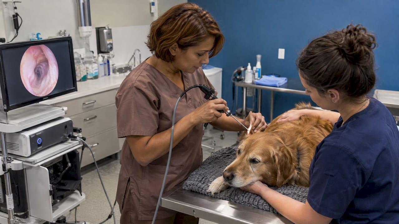
Otoscopy with a rigid endoscope means passing a small telescope, usually 1.9 to 4 mm across, down the ear canal. The telescope is attached to a light source and camera, so you examine the canal and tympanic membrane on a monitor. It gives you more light, more magnification and a working channel for lavage and instruments than a handheld otoscope. For most small animal clinics, a 2.7 mm, 0° rigid telescope is the practical starting point. This guide covers what the technique shows, how to choose optics, how to run the exam, and how to keep it safe.
TL;DR
- A rigid endoscope gives bright distal-tip light, magnification and image capture. Together with a working channel, this makes it a strong step up from a handheld otoscope for deep canal and tympanic work.
- Telescope outer diameters in veterinary use run from 1.9 to 10 mm. For ear work, 1.9 mm to about 4 mm covers most patients, and 2.7 mm is the common all-rounder.
- 0° optics are the routine choice. Angled telescopes widen the view but call for more experience.
- Good technique means aligning the canal by manipulating the pinna, advancing under direct vision, and keeping light power moderate.
- A clean setup (telescope, sheath, light source, camera, monitor) also serves reptile, exotic and other small-diameter work.
Table of Contents
- What rigid endoscope otoscopy actually is
- Why use a rigid endoscope instead of a handheld otoscope
- Choosing the telescope: diameter, angle and length
- The rest of the system
- How to perform the exam, step by step
- Using the endoscope safely
- What the veterinary evidence shows
- Endoscope and microscope: complementary views
- Our perspective
- Equip your clinic for rigid otoscopy
- FAQ
- Sources
What rigid endoscope otoscopy actually is
Otoendoscopy uses a rigid or flexible endoscope to inspect the outer ear, middle ear, mastoid or lateral skull base. Clinically, it can show the tympanic membrane, or the middle ear through a perforation, and can assess residual disease in the operating room.
In veterinary practice the term usually covers two things:
- Diagnostic video-otoscopy. You examine the horizontal canal and eardrum in a dog or cat with magnification, then record images for the chart.
- Therapeutic otoscopy. You flush the canal, remove debris or foreign material, or take a biopsy through the working channel or sheath.
The telescope is a rod-lens design. A light cable from the light source connects at the side, and a camera head sits on the eyepiece. The image goes to a monitor, which lets you, a technician and the owner see the same view.
Why use a rigid endoscope instead of a handheld otoscope
A handheld otoscope is a fine screening tool. The question is what happens when the canal is narrow, painful, swollen or full of debris. A review of veterinary video-otoscopy found it provides greater lighting, magnification, lavage and instrument access than a handheld otoscope. It also helps with documentation and client education.
A 2021 review of endoscopy in otology adds several practical points. Endoscopes provide distal-tip illumination and image capture, a wide-angle view and enhanced depth of field. They also allow access without a straight line of sight, through narrow or tortuous canals.
A 67-dog study of canine otitis externa shows what that means in practice. Every treated dog had an external tympanic concavity holding adherent hair and viscous debris. Direct, magnified viewing close to the tympanum can reveal findings a conventional handheld exam may miss.
A separate review notes that a handheld otoscope is appropriate in mild disease but not when inflammation or debris is heavy. Those are the cases where the endoscope earns its place.

When the exam is indicated
Veterinary video-otoscopy is indicated for pruritus, pain, head shaking, odor, discharge, facial excoriations and aural hematoma. The same source recommends a complete history, physical exam, otic exam, otoscopy and dermatologic evaluation, so the endoscope is one step in a workup and not a replacement for it.
Imaging matters as well. When the tympanic membrane is thickened, hyperemic, absent or bulging, when the malleus is not apparent, or when fluid or a mass is visible behind the membrane, the same review advises assessing the tympanic cavity, ideally with CT or MRI. That helps you decide whether to sample it and reduces risk to a normal middle ear.
Choosing the telescope: diameter, angle and length
Diameter
Veterinary endoscope guidance lists telescope outer diameters from 1.9 to 10 mm and lengths from 10 to 35 cm. For ears, the working range is much narrower.
| Diameter | Typical role in ear work |
|---|---|
| 1.7 to 1.9 mm | Fine inspection of deep or narrow canals, retraction pockets, tympanic cavity work |
| 2.7 mm | The versatile veterinary telescope for otoscopy; its operating sheath can take a 5-Fr instrument such as biopsy forceps |
| 3 to 4 mm | Larger canals and surgical settings; image quality improves as diameter grows |
The 2.7 mm telescope is identified as a common, versatile veterinary telescope for otoscopy. In human office otoendoscopy, 2.7 mm by 11 cm and 1.9 mm by 10 cm rods are described as practical sizes that balance field of view, image quality and access. The same logic applies to a cat or a small dog.
Diameter also trades off against image quality. An otologic safety review notes that common ear-surgery diameters are 2.7, 3 and 4 mm, and that image quality improves as the diameter expands. Pick the largest telescope that passes comfortably. Our guides on choosing the right small animal endoscope size and the endoscope diameter guide walk through that decision by species and body size.
Viewing angle
Telescopes are made with viewing angles from 0° to 120°, but angles above 30° are rarely used in veterinary medicine. Fine rigid endoscopes used to assess retraction pockets in human patients come in 1.9 mm versions with 0°, 30° or 70° views, and 1.7 mm versions with 0°, 30° or 90° views.
For routine canal work, 0° is the safe default. An office otoendoscopy guide says 0° telescopes are safest for routine use. It adds that 30° or 45° may be needed for selected cases, but angled scopes belong in the hands of experienced users because they raise the chance of touching the sensitive bony canal.
Angled optics have a real payoff when used well. They allow views around corners, beyond a microscope's line of sight, into hidden regions such as the retrotympanum and hypotympanum.
What a purpose-built veterinary otoscope looks like
A BSAVA chapter on rigid otoendoscopy describes a small-animal otoscope 8.5 cm long, with a 5 mm tip, an integrated 5-Fr (1.65 mm) working channel, a 110° field of view and a long focal length meant for deep canal examination. Think of the combination as the target: a short, rigid, bright instrument that carries suction, lavage or forceps without a second hand on the canal.
You can browse rigid scopes, arthroscopes and otoscopes in our veterinary rigid endoscopy catalog.
The rest of the system
A rigid ear exam needs more than the telescope.
- Light source. Xenon, LED and halogen sources are all available, and the literature gives no definitive evidence favoring one. Our overview on choosing the best light source for veterinary endoscopy covers the practical trade-offs.
- Camera and monitor. Essential equipment for endoscopic middle-ear surgery includes the light source, 0° and 30° rigid endoscopes, and an HD 3-CCD camera with monitor. Office setups use an HD or 4K camera and monitor, and recording is what turns the exam into a chart entry and a client conversation.
- Sheath and working channel. Suction, lavage and 3- or 5-Fr instruments go through the channel or sheath. A flexible biopsy forceps fits that role, and you can see ours on the biopsy forceps page.
- Lavage supplies. In the canine study, hair and debris were removed with 3- or 5-Fr grasping forceps, and the canal was washed with a 3- or 4-Fr catheter, under more than 10× monitor magnification.
Small-diameter scopes pay off beyond ears. The same 2.7 mm telescope is the usual starting point for exotics, as we explain in our post on why vets start reptile endoscopy with a 2.7 mm scope. More on the wider case is in our piece on small diameter endoscopes in veterinary practice.
How to perform the exam, step by step
The BSAVA chapter's sequence is a sound framework: choose a diameter that fits comfortably, stabilize patient and examiner, align the canal by manipulating the pinna, and advance under direct vision, never by force. Veterinary exams may be performed under general anesthesia with intubation and eye protection, followed by complete removal of lavage fluid.
- Prepare and white-balance. Check the telescope, sheath, light cable and camera. Make sure suction and lavage are connected before the scope goes in the ear.
- Position and protect. Place the patient so the head is stable. Protect the eyes and intubate if the exam is under anesthesia.
- Align the canal. The dog's external canal is about 5 to 10 cm long and 4 to 5 mm wide, with vertical and horizontal portions, so the pinna must be manipulated to line them up.
- Enter at the intertragic incisure. Grasp the base of the pinna and retract it dorsally and slightly laterally to pass the dorsal fold. Then retract laterally and ventrally as you advance through the horizontal canal, as Angus describes.
- Advance only with a clear view. If the image is lost to debris, stop, flush and suction. Do not push through.
- Inspect systematically. The whole canal and tympanic membrane may not be visible at once. A British Society of Audiology procedure advises inspecting portions in turn by gently angling the instrument or changing head position.
- Record. Capture stills or video of the canal, membrane and any lesion before you intervene, and again afterward.
- Treat or sample. Use the working channel for lavage, debris removal or biopsy as indicated.
- Finish clean. Remove all lavage fluid, then reprocess the telescope and sheath according to the manufacturer's instructions.
Our small animal endoscopy tips cover handling habits that carry over to ear work, and the videos of our PortaScopes in use show operation and cleaning procedures.
Using the endoscope safely
Safe use is mostly about light, contact and stability. An office otoendoscopy guide names discomfort and thermal injury as the main risks. It recommends light power below 50%, a supine patient with the head stabilized, and the scope stabilized on the posterior meatus. It also notes that 2D endoscopy relies on motion parallax for depth perception, so slow, deliberate movement helps you judge distance.
On heat, an endoscopic ear surgery review points to possible light-related temperature rise. A cited animal study found the greatest increase with a xenon source and a 4 mm scope. The smallest was with an LED source and a 3 mm scope (Transcanal endoscopic stapedotomy). Keep the lamp as low as the image allows and avoid resting the tip against tissue with the light at full output.
The British Society of Audiology's 2022 procedure is written for human audiology, but its principles carry over. It advises a stable position, bracing the instrument against the head, choosing the largest comfortable tip, and taking particular care because the bony canal is sensitive and its skin vulnerable to trauma.
Finally, one-handed work and loss of stereopsis are known features of endoscopic ear technique. A working channel or a technician holding the pinna and steadying the head helps manage both.

What the veterinary evidence shows
The canine data is encouraging for clinics adopting this technique.
- In the 67-dog otitis externa study, anesthetized dogs were treated with video otoscopy at more than 10× monitor magnification, with hair and debris removed by forceps and the canal washed by catheter (J Vet Med Sci, 2011).
- In a canine cadaver study of 10 total ear canal ablation and lateral bulla osteotomy specimens, a 1.9 mm rigid 30° endoscope found epithelial remnants in at least one of five tympanic cavity regions after every procedure. The rostral region held the highest median proportion (35.6%) and the medial region the lowest (1.8%).
- The authors concluded the 1.9 mm endoscope was effective for evaluating all tympanic cavity sections after curettage, and that it should be considered to improve epithelial removal in initial or revision surgery.
The point for your clinic: visibility inside the canal and bulla is where outcomes can improve, and a small rigid scope is the tool that gets you there.
Endoscope and microscope: complementary views
Ear surgeons often describe the two as complementary. The endoscope gives wide-angle, around-the-corner access and distal illumination. Microscopic surgery keeps its advantages for stereoscopic, two-handed work. Clinics that do both may want to look at microscopes and microscopy cameras alongside their endoscopy setup, since magnified visualization on the bench and in the ear work together.
Our perspective
Most clinics we talk to start ear endoscopy for the same reason: a handheld otoscope stopped answering the question in front of them. A painful, narrow or debris-filled canal needs light, magnification and a way to flush at the same time.
We build our approach around small-diameter rigid optics because they fit the patients veterinarians actually see, from cats and small dogs to exotics. A 2.7 mm telescope with a sheath is a versatile first purchase, and it does not tie up a whole hospital system's budget. Portable systems let the same equipment move between the exam room, the surgical suite and the field.
Our goal is affordable, veterinary-specific equipment with fast shipping and round-the-clock support, so a clinic can start recording ear exams and treating with confidence. The monitor view also changes the client conversation. Owners see the canal themselves, and treatment plans become easier to explain.
Endoscope
Equip your clinic for rigid otoscopy
We supply veterinary rigid scopes, arthroscopes and otoscopes, light sources, flexible biopsy forceps and other endoscopic accessories, along with endoscope repairs. Start with the veterinary rigid endoscopy catalog for telescopes, or see the full range on 1800endoscope.com.
If you are comparing sizes for a mixed caseload, our veterinary endoscope buying guide is a good next read. For documenting findings and sharing them with colleagues, see our article on remote visual inspection techniques for veterinary diagnostics.
FAQ
What size rigid endoscope is best for ear exams in dogs and cats?
A 2.7 mm telescope is the common, versatile veterinary choice for otoscopy, and its sheath can take a 5-Fr instrument such as biopsy forceps. For narrow canals or tympanic cavity work, 1.9 mm telescopes are used. Match the diameter to the patient's canal.
Do I need anesthesia for rigid endoscope otoscopy?
Veterinary exams are often performed under general anesthesia with intubation and eye protection, particularly when you flush, remove debris or sample. Anesthesia keeps the patient still and the canal aligned. Your clinical judgment and the patient's condition decide.
Is a 0° or 30° telescope better?
For routine ear exams, 0° is the safest and most common choice. Angled scopes show more around corners but carry a higher risk of contacting the bony canal, so reserve them for experienced users and selected cases. Angles above 30° are rarely used in veterinary medicine.
What else do I need besides the telescope?
You need a light source and cable, a camera and monitor, and a sheath with a working channel for lavage, suction and instruments. Lavage catheters and small grasping or biopsy forceps complete the setup. Recording capability helps with charting and client education.
Sources
- Basic principles of endoscopic ear surgery
- Advances in Otoscopy
- Current trends and applications in endoscopy for otology
- Treatment of Canine Otitis Externa Using Video Otoscopy
- Rigid endoscopy: otoendoscopy, BSAVA Library
- Endoscopic Equipment for the Veterinary Practitioner
- Office-Based Otoendoscopy
- Endoscopic Diagnosis and Follow-up of Retraction Pockets and Cholesteatomas
- Staying safe during endoscopic ear surgery
- Transcanal endoscopic stapedotomy
- Otoscopic evaluation of epithelial remnants in the tympanic cavity
- BSA Recommended Procedure: Ear Examination
- Tienda Microscopios