
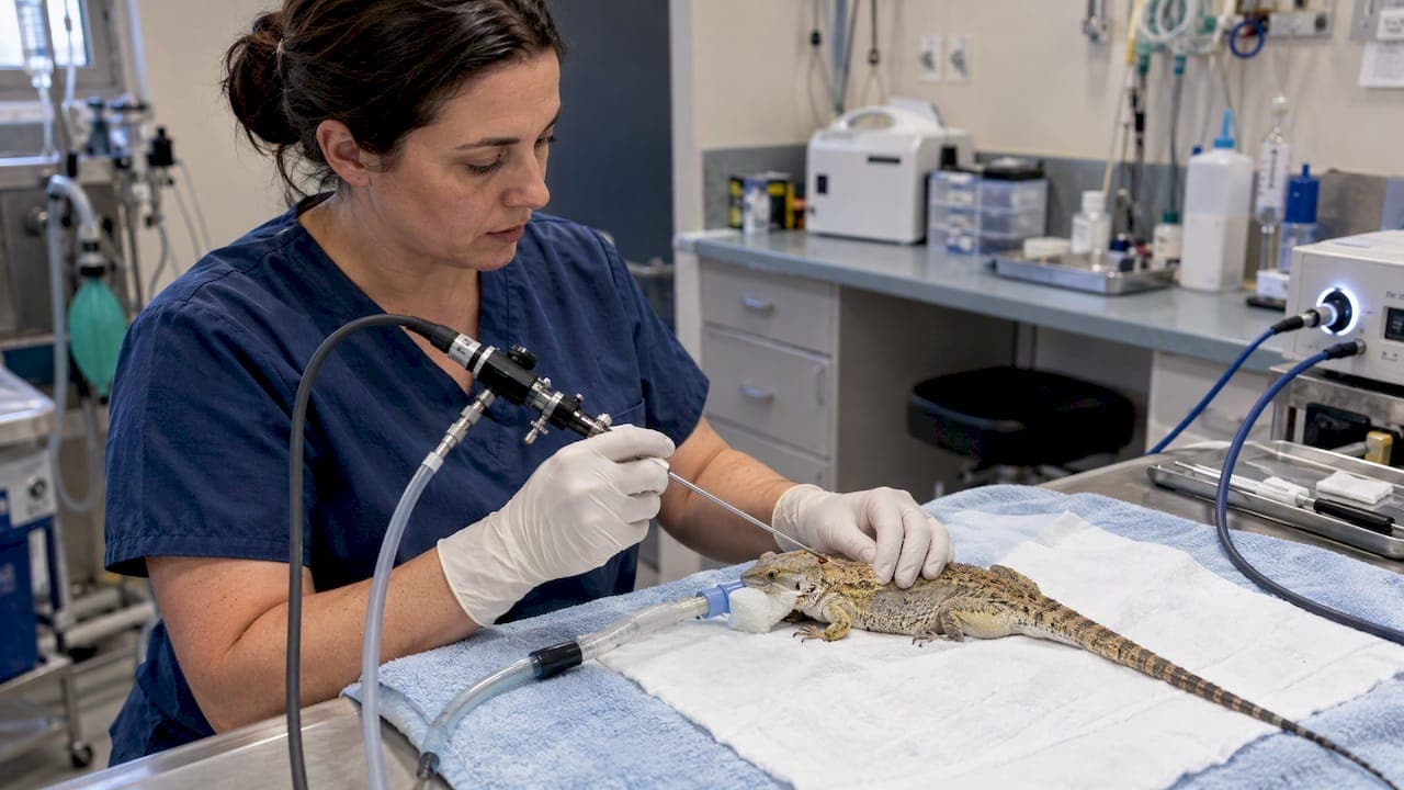
A reptile endoscopy setup needs several things to function in daily practice: a rigid or flexible scope, a light source and camera, a monitor, biopsy forceps with a matching operating sheath, and insufflation or irrigation accessories alongside proper reprocessing supplies. The rule that governs every purchase is simple: pick the smallest-diameter scope that still accommodates the instruments and visualization a procedure demands, and a 2.7 mm system is usually the most practical starting point. Every invasive procedure also requires general anesthesia and reprocessing that follows the manufacturer's instructions for use.
TL;DR:
- A 1.9 mm scope suits smaller patients and diagnostic exams without a working channel; a 2.7 mm system typically accommodates 1.7 mm biopsy forceps.
- Rigid optics generally provide brighter, sharper images for coelioscopy and biopsies, while flexible scopes suit airway work and selected gastric procedures.
- Clinicians must stabilize sick patients, monitor heart rate and temperature, and set criteria for converting invasive procedures under general anesthesia to open surgery.
- After each case, follow the scope manufacturer’s instructions for precleaning, leak testing, brushing, and disinfection; biopsy forceps that breach tissue require sterilization.
Table of Contents
- Rigid, flexible, and video options for reptile patients
- Matching scope diameter to species and procedure
- Building the rest of the system: light, camera, and accessories
- Anesthesia and monitoring during the procedure
- Reprocessing and infection control between cases
- Training and technique that reduce procedural risk
- Why we favor versatile, cost-conscious equipment choices
- Finding the right starting kit for your clinic
- FAQ
- Sources
Rigid, flexible, and video options for reptile patients
Rigid endoscopes pair a telescope with an outer sheath, giving a fixed, stable line of sight and the option to add an instrument channel for forceps or irrigation. Flexible endoscopes bend around anatomy, which matters for airway and some gastrointestinal work where a straight approach will not reach the target.
Within both families, the image comes from either a videoscope with a camera chip built into the tip, or a fiber-optic bundle feeding an external camera head. Rigid telescopes generally deliver sharper, brighter images and steadier handling, which is why they dominate coelioscopy and liver or kidney biopsy in lizards and snakes. Flexible scopes earn their place in bronchoscopy and certain gastric procedures where reach outweighs image quality, though flexible units in small diameters tend to show more pix elation than rigid equivalents of the same size. A 2.7-mm rigid system has been used safely for coelioscopy and liver biopsy across a range of lizard species, with short anesthesia times and biopsy samples adequate for histology. For most clinics building a first reptile endoscopy setup, that case history makes rigid optics the sensible anchor, with a flexible scope added later for airway cases.

Matching scope diameter to species and procedure
Diameter determines what a scope can see and what instruments can pass alongside it, so sizing comes before any other purchasing decision. The most common working diameters in reptile practice run from 1.9 mm and 2.7 mm up to 3 mm, with larger 5 to 10 mm scopes reserved for sizeable chelonians or open surgical endoscopy. Rigid telescopes in the 1.9 to 2.7 mm range are versatile enough for most exotic mammal and reptile work, and many of the instruments used with them are adapted from pediatric or ophthalmic sets.
Working channel size sets a hard ceiling on instrument choice: a 2.7 mm system typically pairs with roughly 1.7 mm biopsy forceps, while a 1.9 mm scope without a channel is best suited to diagnostic exams unless used alongside a separate access port. The practical decision rule looks like this:
- A 2.7 mm rigid system covers coelioscopy, liver and kidney biopsy, and general diagnostic exams in most pet lizards and snakes.
- A 1.9 mm scope suits smaller patients or spaces where a channel is not required for the exam.
- A larger 5 to 10 mm scope fits big chelonians, cystoscopy in larger animals, or cases converting toward surgical endoscopy.
A clinic seeing mostly bearded dragons, ball pythons, and similar-sized patients rarely needs anything beyond the 2.7 mm tier to start.
Building the rest of the system: light, camera, and accessories
The scope is only one piece of a working setup. A light source, either LED or xenon, drives image brightness and color accuracy, and it needs to match the scope's fiber or light-post connection. The camera processor converts the optical image into video, with options ranging from basic fiber-optic couplers to full HD videoscopes, and a monitor displays the feed in real time. Recording capability, whether SD-card storage, HD output, or wireless transmission to a tablet, matters for case documentation and client communication.
Beyond imaging, a functioning system needs insufflation and irrigation pumps to maintain a working space inside the coelomic cavity, along with valves, suction adapters, and an operating sheath sized to the telescope. On the instrument side, biopsy forceps, retrieval (grasping) forceps, and cleaning adapters round out a basic kit. Smaller practical additions, such as a scope-holding arm or magnifying loupes, reduce hand fatigue during longer procedures and help steady the image in small patients.
Anesthesia and monitoring during the procedure
Invasive reptile endoscopy, including coelioscopy, requires general anesthesia rather than sedation alone; some brief, non-invasive exams may tolerate sedation, but complete immobilization is preferred whenever instruments enter a body cavity. Isoflurane and ketamine are the agents most frequently cited as adequate for these procedures.
Stabilizing a sick reptile before induction often matters more than the procedure itself, and clinicians should be ready to delay, abort, or convert to open surgery if the patient's condition shifts under anesthesia. Monitoring in small ectotherms leans on practical tools: a Doppler probe for heart rate, active warming to maintain body temperature, and capnography or pulse oximetry where the equipment and patient size allow. Keeping procedure times short reduces anesthetic risk in patients that often weigh under a kilogram.
Reprocessing and infection control between cases
Reprocessing is not optional paperwork, it is what keeps a scope safe to reuse. The accepted sequence starts with point-of-use pre-cleaning immediately after the procedure, followed by leak testing, manual brushing and flushing of channels, and then high-level disinfection or sterilization according to the manufacturer's instructions for use. Automated endoscope reprocessors are strongly recommended over manual HLD alone because they deliver more consistent cycles and easier documentation.

Reprocessing guidance modeled on SGNA and FDA standards calls for checking that any AER is compatible with the specific scope model before use, and for classifying each accessory as critical or semi-critical. Items that breach mucosa or enter sterile cavities, such as biopsy forceps, need full sterilization rather than HLD. Our step-by-step sterilization walkthrough and 2026 reprocessing guide cover the adapters and documentation a clinic needs to keep this process audit-ready.
Training and technique that reduce procedural risk
Reading equipment specifications only goes so far; hands-on practice is what builds the muscle memory for safe scope handling. Continuing-education courses such as the exotic endoscopy program at the University of Georgia pair didactic sessions with supervised case work on anesthetic risk management and endosurgical technique.
Pro Tip: Support the scope with your dominant hand at the camera-sheath junction and steer the tip with the other hand. This two-hand method cuts tremor noticeably in patients under 1 kg.
Start with a single versatile 2.7 mm system, then add instrumentation as caseload grows, and set clear thresholds in advance for when a case converts to open surgery rather than pushing an endoscopic approach too far. Our small-animal endoscopy technique tips walk through handling adjustments for exotic patients specifically.
Why we favor versatile, cost-conscious equipment choices
We stock veterinary endoscopes and related equipment for clinical use. Our stance is practical: verify IFU compatibility before you buy anything, and request a repair quote before committing to a full build-out. Starting with a 2.7 mm rigid system and expanding as your reptile caseload grows keeps the initial investment proportional to actual demand.
— Endoscope
Finding the right starting kit for your clinic
Matching a reptile endoscopy setup to your caseload comes down to picking the right starting category rather than buying every accessory at once. Our veterinary rigid endoscopy catalog covers rigid endoscopy equipment for veterinary practices. For airway inspection or portable field work, our borescope and videoscope line offers videoscopes.
Instrument selection follows the same logic: our flexible biopsy forceps are available for endoscopic procedures. For clinics expanding into broader endoscopic and surgical accessory needs, our medical endoscope and accessories page includes endoscopes and accessories.
If you already own equipment that needs service, our repair and core equipment page is the place to start a repair request or ask about parts availability. Whichever path fits your practice, reach out for an IFU compatibility check before your next order.
FAQ
What is the best scope diameter for reptile endoscopy?
A 2.7 mm rigid system is the most versatile starting point for pet lizards and snakes, supporting coelioscopy, biopsy, and general diagnostics in most reptile species. Larger chelonians or surgical cases may call for scopes in the 5 to 10 mm range instead.
Do reptiles need general anesthesia for endoscopy?
Invasive procedures like coelioscopy require general anesthesia for complete immobilization rather than sedation alone. Isoflurane and ketamine are the agents most commonly cited as adequate for these cases.
How should reptile endoscopy equipment be sterilized between patients?
Reprocessing follows manufacturer IFU and includes point-of-use pre-cleaning, leak testing, manual brushing, and then high-level disinfection or sterilization. Accessories that breach mucosa, such as biopsy forceps, require full sterilization rather than HLD.
Where can veterinarians get hands-on reptile endoscopy training?
Continuing-education programs such as the exotic endoscopy course at the University of Georgia combine lecture content with supervised handling practice. Pairing this training with early cases under a 2.7 mm system builds technique before a clinic takes on more complex procedures.
What accessories does a basic reptile endoscopy setup require?
A working setup needs a light source, camera processor, monitor, biopsy and retrieval forceps, and an operating sheath sized to the scope's working channel. Insufflation and irrigation pumps round out the kit for coelioscopy and similar procedures, and our rigid endoscopy catalog lists rigid endoscopy equipment.
Sources
- Depts
- Ovid
- Endoscopic examination of the pleuro-peritoneal cavity in reptiles
- 2026 Avian, Reptile, Small Mammal, and Fish Endoscopy (Online Course) - University of Georgia College of Veterinary Medicine
- STANDARDS FOR INFECTION CONTROL AND REPROCESSING OF FLEXIBLE GASTROINTESTINAL ENDOSCOPES