
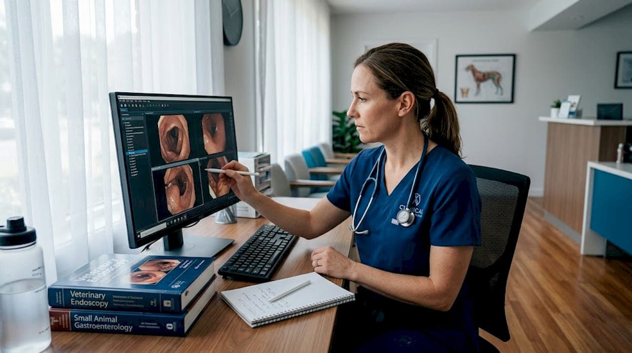
Most veterinary professionals assume that accurate diagnostics require the animal right in front of them. That assumption made sense before modern imaging technology changed the game. Remote visual inspection (RVI) is the process of examining physical conditions from a distance using digital capture and a separate review workflow, typically when direct access is unsafe, impractical, or logistically complicated. In veterinary practice, RVI is reshaping how clinicians examine airways, gastrointestinal tracts, reproductive systems, and oral cavities, especially when specialist expertise is miles away or the animal cannot be safely transported.
Table of Contents
- What is remote visual inspection?
- Veterinary use cases and RVI technology
- Key requirements and constraints for remote inspection
- Clinical effectiveness and risk management
- A practical perspective: What most guides miss about remote visual inspection in animal health
- Explore advanced RVI systems and solutions
- Frequently asked questions
Key Takeaways
| Point | Details |
|---|---|
| Essential workflow | Remote visual inspection uses imaging and remote analysis to expand access to animal diagnostics. |
| Veterinary applications | RVI enables remote airway, GI, dental, and internal examinations for quick clinical decisions. |
| Technology matters | The quality of endoscopes, video probes, internet, and lighting controls RVI effectiveness. |
| Know the limits | RVI can't replace all physical exam components; escalation protocols are vital for safety. |
| Risk management | Remote triage aligns with in-clinic results most of the time, but protocols reduce under-triage risks. |
What is remote visual inspection?
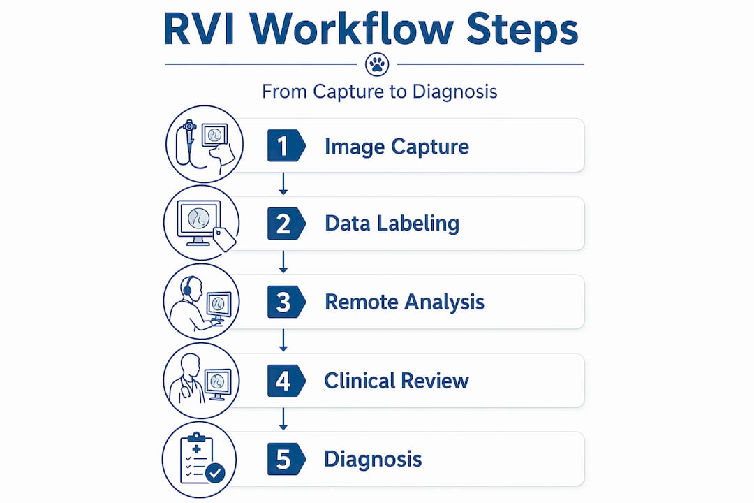
RVI is not a single device or product. It is a structured workflow that connects high-quality image or video capture at the animal's location to expert analysis that can happen elsewhere. The three core stages are data acquisition, remote analysis, and clinical documentation.
In the data acquisition stage, an on-site technician or veterinarian uses a specialized imaging device, typically an endoscope, borescope, or video probe, to capture real-time footage of the target anatomy. That footage is then transmitted digitally to an off-site specialist, a university teaching hospital, or a senior clinician reviewing the case on a tablet or workstation. Finally, the findings are documented in the patient record with annotated images or timestamped video clips.
This workflow directly mirrors how remote visual inspection works in industrial settings, where cameras and optical instruments such as borescopes and video probes allow experts to evaluate hard-to-reach internal surfaces without being physically present. The underlying principles translate cleanly to veterinary medicine.
| RVI stage | What happens | Key tool |
|---|---|---|
| Data acquisition | Image or video captured at patient location | Endoscope, borescope, video probe |
| Remote analysis | Specialist reviews footage off-site | Secure video platform, cloud storage |
| Clinical documentation | Annotated findings entered into patient record | EMR software, image management system |
The key advantages RVI offers over traditional examination include:
- Access to anatomy that is difficult or impossible to visualize without instrumentation
- Specialist consultation without requiring animal transport
- Objective, reviewable documentation for ongoing case management
- Reduced procedure time and patient stress in straightforward cases
"RVI is not just a technology upgrade. It is a rethinking of where clinical expertise needs to be at the moment of examination."
For a broader overview of how endoscopic technology supports these workflows, the veterinary endoscopy overview at 1800endoscope.com covers the full equipment ecosystem clearly and practically.
Veterinary use cases and RVI technology
Understanding the basics puts you in a position to apply them. Across small animal, equine, and large animal practice, RVI tools are addressing diagnostic gaps that were previously handled either with referral or educated guesswork.
Common veterinary RVI applications by species and anatomy:
-
Airway and respiratory evaluation in horses and dogs: Flexible endoscopes pass through the nasal passage or oral cavity to visualize the larynx, trachea, and bronchi. In horses, this is essential for diagnosing exercise-induced pulmonary hemorrhage, recurrent laryngeal neuropathy, and dorsal displacement of the soft palate.
-
Gastrointestinal examination in companion animals: Small-diameter flexible scopes allow visualization of the esophagus, stomach, and proximal small intestine without invasive surgery. Gastric foreign bodies, ulcers, and mucosal lesions become visible in real time.
-
Reproductive tract evaluation in livestock: Video probes are used in large-scale production settings to assess reproductive health without the logistical challenge of moving animals to a clinic.
-
Dental and oral cavity inspection: Compact videoscopes capture high-resolution images of tooth roots, gingival pockets, and oral lesions in cats, dogs, and exotic species where direct visualization is nearly impossible without magnification.
-
Ear canal examination: Otoscopy-style video devices document debris, polyps, and eardrum integrity in a format that can be shared with dermatologists or specialists for remote second opinions.
These applications rely on hardware modalities including endoscopes, borescopes, and video probes. As industrial RVI principles applied to veterinary diagnostics demonstrate, the guidance and interpretation protocols transfer effectively once you map acceptance criteria to veterinary diagnostic standards.
| Device type | Best veterinary use | Key feature |
|---|---|---|
| Flexible videoscope | GI tract, airway, upper respiratory | Steerable tip, HD video output |
| Rigid endoscope | Joint, bladder, nasal cavity | High image clarity, small diameter |
| Portable borescope | Field examination, barn-side consults | Battery-operated, wireless streaming |
| Dental videoscope | Oral cavity, ear canal | Compact head, macro imaging capability |
The portable airway inspection endoscope is a strong example of field-ready hardware designed for equine respiratory assessment. For barn-side consultations with remote specialists, an equine field scope with USB connectivity enables real-time streaming to off-site clinicians. For oral and dental work, a dental videoscope provides the magnification and documentation needed for referral-quality records.
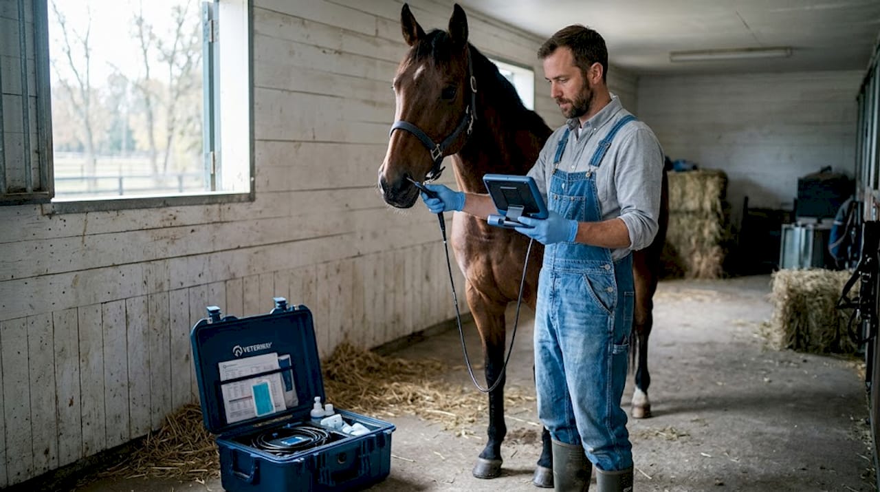
Pro Tip: Standardize your image metadata before any RVI session. Label each file with the animal ID, anatomy examined, date, device used, and the name of the reviewing clinician. This one habit dramatically speeds up specialist review and strengthens your medical record.
Key requirements and constraints for remote inspection
Making RVI work consistently requires more than good equipment. There are technical and procedural prerequisites that determine whether the remote review produces actionable clinical conclusions.
Critical requirements for reliable RVI in veterinary settings:
- High-resolution imaging hardware: Blurry or low-contrast footage defeats the purpose. Resolution matters most in mucosal tissue assessment, where subtle color changes or surface texture differences indicate pathology.
- Adequate lighting: Endoscopic light sources must be powerful enough to illuminate deep anatomical structures without washout. This is where purpose-built veterinary scopes outperform improvised solutions significantly.
- Reliable data transfer: Whether you are streaming live or transmitting recorded footage, connectivity needs to be stable. A dropped connection during a key moment in the examination creates gaps in the record and delays diagnosis.
- Structured examination protocol: The on-site operator must follow a defined sequence so the reviewing specialist sees all relevant anatomy, not just what the technician found interesting.
- Trained operators: A high-quality scope in untrained hands still produces poor data. Staff training on scope handling, patient positioning, and image capture directly impacts the quality of the remote review.
Remote veterinary workflows that succeed consistently cite video quality and adequate lighting as the most critical prerequisites, not the technology brand or the specialist's credentials. Those factors matter too, but they cannot compensate for poor visual data.
"Visualization constraints can become the limiting factor more than clinician expertise alone when imaging prerequisites are not met."
It is also important to acknowledge what RVI cannot do. Palpation, auscultation, neurologic reflex testing, and tactile assessment are outside the scope of any visual-only workflow. A limping horse, a dog with an irregular heartbeat, or a cow showing vague signs of systemic illness may require hands-on evaluation that no camera system can substitute.
For answers to common technical questions about endoscope selection and compatibility, the endoscope FAQ provides straightforward guidance organized by clinical application.
Pro Tip: Before any field RVI session, particularly emergency or remote barn calls, test your imaging system and internet connection end-to-end. Run a brief mock capture and transmission so you know the setup works before the animal is in position.
Clinical effectiveness and risk management
RVI performs well in defined diagnostic scenarios, but it is not without meaningful limitations that demand structured risk management.
Veterinary telemedicine research reports approximately 70% concordance between remote triage urgency assessments and subsequent in-clinic determinations. That figure is encouraging but it also means roughly 30% of cases show some level of discordance. More critically, under-triage occurs in 10 to 15% of cases when remote video-based review is conducted without clear escalation criteria. Under-triage means a case that needed urgent care was assessed as lower priority, with obvious implications for animal welfare.
Statistical callout: Up to 15% of remotely triaged veterinary cases may be under-triaged without written escalation protocols in place. That figure should drive every clinical team to formalize their RVI safety framework before routine deployment.
A practical risk management checklist for RVI workflows:
-
Define the case scope. Specify which case types are suitable for RVI review and which require mandatory in-person evaluation. Create a written list and update it as your team gains experience.
-
Assign escalation criteria. Document exactly when a remote case must be escalated to physical examination. Include red-flag signs visible on video as well as owner-reported symptoms that automatically trigger in-person triage.
-
Require dual confirmation for high-stakes findings. Any remote diagnosis with significant treatment implications, including suspected foreign body, mass, or acute hemorrhage, should be confirmed by a second reviewer or by in-person follow-up.
-
Log every RVI session formally. Time-stamp records, device used, reviewer name, findings, and follow-up recommendations. This documentation supports quality review and legal protection.
-
Review outcomes regularly. Track cases where RVI diagnosis was later revised after in-person examination. Analyze patterns to identify where your protocols need adjustment.
Signs that a case should escalate immediately from RVI to physical evaluation:
- Abnormal sounds or breathing patterns that the video cannot characterize adequately
- Unexplained weight loss or systemic signs without a clear visual finding
- Neurologic symptoms requiring reflex testing
- Owner-reported rapid deterioration between video capture and specialist review
- Any finding where treatment risk is high and visual data alone cannot rule out alternatives
For cases where definitive diagnosis requires hands-on instrumentation, veterinary rigid endoscopes provide the direct visualization and procedural access that remote review cannot replicate.
A practical perspective: What most guides miss about remote visual inspection in animal health
Most RVI content for veterinary professionals focuses on the device. Resolution specs, insertion tube diameter, wireless connectivity, battery life. These details matter, but they are not where RVI programs succeed or fail in real clinical environments.
Here is what actually determines whether an RVI workflow delivers value: the quality of the protocol wrapped around the device. We have seen clinics invest in excellent endoscopic hardware and still produce inconsistent results because nobody defined how images should be labeled, who reviews them, how long review should take, or what happens when a finding is ambiguous. The device is the easy part.
Staff training is systematically underestimated. A skilled endoscopist produces footage that a specialist can confidently interpret in minutes. An untrained operator using the same device produces footage that raises more questions than it answers. Investing in training is not optional if you want RVI to function as a genuine diagnostic extension rather than an expensive documentation tool.
Standardizing metadata is equally undervalued. When a specialist receives an unlabeled video clip from a field location, they have to spend time gathering context before they can focus on the clinical question. When every file arrives with animal ID, date, anatomical region, device type, and relevant clinical history embedded in the filename or attached form, that specialist can move directly to interpretation. The time savings across a busy referral practice are significant.
Perhaps the most important mindset shift is treating RVI as a strategic extension of hands-on care, not as a replacement for it. Clinics that deploy RVI most effectively use it to extend specialist access into field settings, accelerate triage decisions for stable cases, and build richer documentation for chronic disease management. They do not use it to avoid necessary physical examinations.
Write your escalation protocol before your first remote session. It is tempting to build this iteratively once you know what cases come your way, but the under-triage risk is highest when the program is new and protocols have not yet been tested. Define the boundaries first, then refine them with experience.
Transparent documentation and structured feedback loops are what separate programs that improve over time from those that plateau. Every case where a remote finding differed from the in-person result is a learning opportunity. Capture those cases systematically, review them as a team, and update your protocols accordingly.
Explore the full range of veterinary endoscopy systems to understand which platforms are best suited to supporting a structured RVI workflow in your specific practice environment.
Explore advanced RVI systems and solutions
If you are ready to build or upgrade your remote visual inspection capability, choosing the right equipment for your patient population and field conditions makes a measurable difference in diagnostic confidence.

At 1800endoscope.com, veterinary clinics and animal health professionals can access a full catalog of portable veterinary endoscopes designed for airway, GI, and reproductive examinations across small and large animal species. For procedures requiring rigid access with high image clarity, rigid endoscopes for clinics cover nasal, joint, and bladder applications. The full range of endoscopy solutions includes compatible light sources, recording systems, biopsy accessories, and cleaning tools, so you can build a complete, field-ready RVI system from a single source. Expert consultation is available to match hardware to your specific diagnostic workflow.
Frequently asked questions
How does remote visual inspection improve veterinary diagnostics?
RVI enables clinicians to visually assess internal anatomy from a distance using endoscopic capture and digital transmission, and this expands diagnostic access for cases where transport is impractical or specialist proximity is limited.
What are the main limitations of remote visual inspection in animals?
RVI cannot replicate palpation, auscultation, or neurologic testing, and under-triage failure modes affect approximately 10 to 15% of cases when escalation protocols are absent.
Which remote visual inspection tools are most popular in veterinary applications?
Flexible endoscopes, rigid borescopes, and portable videoscopes are the most widely used platforms, with cameras and optical instruments designed for internal surface visualization leading clinical adoption.
What prerequisites are needed for successful remote visual inspection?
Video quality and adequate lighting are the most consistently cited prerequisites in successful veterinary remote inspection workflows, alongside stable data connectivity and trained on-site operators.
Recommended
- FULLY PORTABLE 6mm X 150 AIRWAY INSPECTION DIRECT MONITOR SD CARD VIDEO AFFORDABLE LOW COST ENDOSCOPE ENDOSCOPY SYSTEM!
- Equine Dental Inpection Videoscope
- EQUINE 8mm USB AIRWAY INSPECTION FIELD SCOPE MONITOR TYPE SD-CARD VIDEOSCOPE
- Medical Veterinary Endoscope Endoscopy Systems, Light Sources, Accessories, Industrial NDT Borescopes