
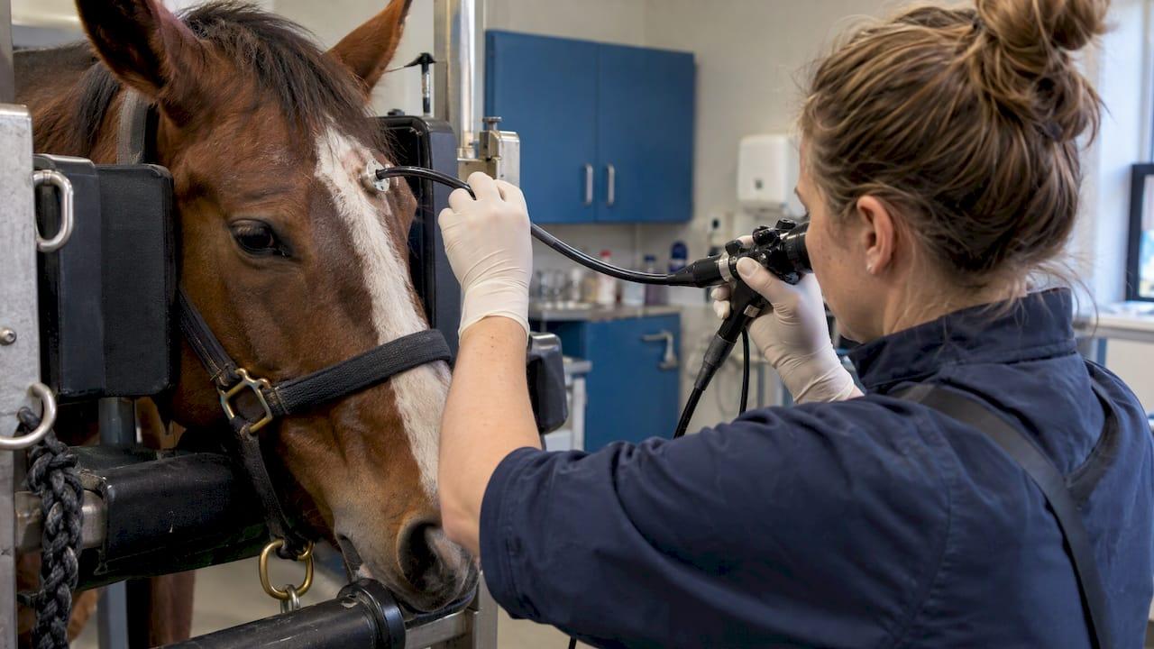
A standing sinoscopy needs a sterile flexible or video endoscope (or an arthroscope), a Galt trephine, local anesthetic, irrigation or suction, and instruments to open the sinus septa when required. It also needs a reprocessing routine that protects the scope between horses. This guide goes through each piece, explains how to choose a scope, and covers recording, cleaning and storage.
TL;DR
- Core kit: sterile flexible fiberoptic or video endoscope (or arthroscope), a 9.5 mm to 15 mm Galt trephine, local anesthetic, and irrigation or suction for exudate.
- Flexible scopes reach most of the paranasal sinus structures with the horse standing. Video endoscopes add better images and recording.
- Rhinoscopy guidance favors scopes of 80 to 120 cm and under 10 mm in diameter.
- Clean with enzymatic detergent right after use, disinfect at the right level, and hang the scope vertically for storage.
- Our equine airway scopes with SD-card or USB recording are listed below for clinics building or upgrading a sinus setup.
Table of Contents
- What sinoscopy is used for
- The equipment list, item by item
- Rigid or flexible: choosing the scope
- Length and diameter: what to check
- Portable setups for barn and field
- Preparing the room and the team
- Where imaging fits alongside sinoscopy
- Cleaning, disinfection and storage
- Perspective
- Build your sinoscopy kit with us
- FAQ
- Sources
What sinoscopy is used for
Sinoscopy is direct visualization of the inside of the paranasal sinuses. The American College of Veterinary Surgeons describes it as making a small opening through the bone over the sinus, inserting a flexible endoscope to evaluate and potentially treat the problem, usually under standing sedation.
It helps with a long list of findings: primary or secondary sinusitis, progressive ethmoid hematoma, sinus cysts, neoplasia, thickened or inflamed mucosa, space-occupying lesions, exudate, dental-associated abscesses and mycotic lesions. In one standing-horse series summarized by DVM360, sinoscopy gave an exact diagnosis in 70% of patients, against 20% for endoscopy and 36% for radiography. A long-term review of 200 cases found sinoscopy was performed in 79% of cases and was of great diagnostic value.
The Merck Veterinary Manual adds that after the trephine hole is made, the scope allows direct inspection of the frontal or maxillary sinuses. The same session can support diagnosis, debridement or removal of purulent debris, and placement of an indwelling lavage system. That is why the equipment list is wider than just a scope: you are diagnosing and treating through one portal.

The equipment list, item by item
The DVM360 standing-horse article lists the working set. Here it is as a checklist, with what each item does.
| Item | Role in the procedure |
|---|---|
| Sterile flexible fiberoptic or video endoscope, or an arthroscope | Visualizes the sinus compartments through the portal |
| 9.5 mm to 15 mm Galt trephine | Creates the opening in the bone over the sinus |
| Local anesthetic | Desensitizes the site, and topical use on the septal bulla |
| Irrigation or suction equipment | Clears exudate that obscures the view |
| Ferris-Smith arthroscopic rongeur, crocodile forceps or diode laser | Fenestrates the maxillary septal bulla through the same portal |
Source for the list: DVM360, Sinoscopy in the standing horse.
Scope
The scope is the decision with the biggest effect on image quality and workflow, so it gets its own section below.
Trephine and anesthesia
The trephine is sized between 9.5 mm and 15 mm. Choose the size to suit your scope and the instruments you plan to pass. Sedation and local anesthesia are used for the standing approach, as the Merck manual describes.
Irrigation and suction
Exudate is common in sinus disease, and it blocks the view quickly. Have irrigation fluid and suction ready before you start rather than hunting for them mid-procedure. Sterile lavage fluid, tubing and a means of draining the sinus should be on the same trolley as the scope.
Instruments for the septal bulla
When the maxillary septal bulla needs opening, DVM360 describes fenestrating it under endoscopic guidance with a rongeur, crocodile forceps or diode laser through the same portal as the endoscope. If bleeding from fenestration obscures the view, the article advises repeating sinoscopy after 24 to 48 hours. Flexible biopsy forceps are part of the same instrument family, and you can browse flexible biopsy forceps and endoscopic accessories on our site.
Rigid or flexible: choosing the scope
A rigid arthroscope can do the job in some cases, but the geometry of the sinuses favors flexible scopes. IVIS notes that most sinus structures can be examined with a flexible video or fiberoptic endoscope in the standing horse, while a rigid arthroscope gives an inferior field of view because it cannot navigate around structures such as the infraorbital canal.
That leaves a second choice: fiberoptic or video.
| Factor | Fiberoptic scope (fibroscope) | Video endoscope |
|---|---|---|
| Image quality | Lower | Superior, with improved illumination |
| Recording | Needs an added camera | Digital images can be transmitted and recorded |
| Field use | Portable and practical | Portable models exist for field work |
| Durability | More susceptible to mechanical damage | Depends on handling and model |
The comparison follows a review in Equine Veterinary Education on rhinoscopy in horses, which notes that fibroscopes cost less and suit field conditions, while videoendoscopes give better images and allow recording.
For a clinic that wants to document lesions, show owners the findings and keep records, video with recording is the practical route. Our guide to optimizing endoscope video recording covers the settings and habits that make saved footage useful.
Length and diameter: what to check
Specifications matter more than brand names. The same Equine Veterinary Education review recommends flexible endoscopes of 80 to 120 cm and 5 mm to 12 mm for rhinoscopy, with under 10 mm preferred for access and maneuverability. Because the nasal cavity sits rostrally, lengths of 80 to 100 cm are described as more efficient.
A published example from Austria shows how slim a working scope can be. Jehle and colleagues used a 5.9 mm pediatric gastroscope, 1 m long, for transnasal procedures in standing sedated horses, applying mepivacaine 2% topically through the working channel. Their paper also notes that minimally invasive transnasal approaches can have an excellent cosmetic outcome, often shorter surgery time, and the potential for outpatient treatment.
When you compare scopes, check these points before you buy:
- Insertion tube length and outer diameter. Match them to the sinus approach you use, not only to general airway work.
- Working channel. If you plan to pass forceps, topical anesthetic or lavage, you need a channel for it.
- Light source and image path. Built-in monitor models are quick to set up in a barn. Check how the light is delivered.
- Recording format. SD card or USB output makes it easy to move footage into the patient record.
- Repair and service. A scope that cannot be repaired locally becomes downtime. We offer endoscope repairs, so ask us about yours.
- Compatibility. Confirm that forceps, valves and adapters fit the scope you choose.
For a wider look at length by use case, see three equine endoscope lengths every clinic should own.
Portable setups for barn and field
Many equine practices do their work away from a hospital. A sinus case may start in the barn with an airway exam and end with a referral or a standing procedure at the clinic. Our equine range is built around portable video scopes with direct monitors and SD-card recording:
- The 8 mm USB airway inspection field scope, a monitor-type SD-card videoscope.
- The 8 mm airway inspection field scope, economy version, also monitor type with SD card.
- The fully portable 9.5 mm airway inspection endoscope system with a direct monitor and SD card.
- The PVS3M semi-portable 3 meter equine endoscope for longer reach work.
- The 12.9 mm by 3.5 meter portable gastroscope with SD card and USB PC output, and the economy 3 meter monitor gastroscope.
Check each product page for diameter, length, channel and accessory details to match your planned technique. Our write-up on equine sinoscopy setup, room and procedure shows how to lay out the stocks, trolley and monitor so the team is not reaching across the horse's head.
Preparing the room and the team
The equipment list is only half the setup. A few habits keep the procedure smooth:
- Position the monitor where the endoscopist can see it without turning away from the horse's head.
- Lay out the sterile scope, trephine, anesthetic and irrigation in the order you will use them.
- Test the light and the recording before sedation, not after.
- Assign one person to watch the monitor and the recording, and another to handle fluids and suction.
- Keep spare accessories, such as a second set of forceps, within reach.
Where airway work is part of the same visit, our piece on equine airway endoscopy equipment can help you plan a kit that serves both jobs.
Where imaging fits alongside sinoscopy
Sinoscopy is one tool in a diagnostic plan. Radiographs of the head can make lesion localization hard because structures overlap, and a Frontiers in Veterinary Science review notes that CT removes that superimposition. The same review describes standing CT as lower risk than general anesthesia, and notes that cone-beam units have limits with patient size, motion and soft tissue image quality.
In practice, imaging maps the disease and sinoscopy lets you see it, sample it and treat it, often in a standing horse. A clinic without CT on site can still build a strong sinus workup around good endoscopic equipment, and refer for CT when the case calls for it.
Cleaning, disinfection and storage
A sinus scope is only as good as the reprocessing behind it. The Equine Veterinary Education review sets out this sequence:
- Clean immediately after use to remove organic residue, with enzymatic detergent on the working channels and on the outside and inside surfaces.
- Rinse with running water and air-dry the working channel.
- If a pathogen with high transmission potential is suspected, sterilize. The review describes 2% glutaraldehyde as the most recommended method.
- Store the scope in a clean, dry place, preferably hung vertically, and never coiled in its case.
An infection control manual from the University of Queensland veterinary school classes endoscopes as semi-critical equipment: high-level disinfection is required in relatively sterile areas, while thorough low-level disinfection is considered adequate in non-sterile areas when a transmissible disease was not suspected in the previous patient. Because a sinoscopy goes through a bone portal into a sinus, plan your protocol with your own infection control policy and your scope manufacturer's instructions.
The same manual notes that chemical sterilants such as glutaraldehyde are typically used for scopes because most cannot be autoclaved, and it advises using a timer to measure the exact contact time, since too little means inadequate killing and too much can damage the instrument.
For detailed routines, see our step-by-step endoscope sterilization guide, the veterinary sterilization overview and our advice on storing endoscopic equipment.
Perspective
We hear the same story from equine practices: the sinus case arrives, and the scope on the shelf was bought for airway checks. It works, but it was not chosen with the sinus in mind.
Our view is that a clinic gets more from a portable video scope with direct recording than from a cheaper setup that cannot capture what the vet sees. Recording turns a procedure into a record you can review, compare at a recheck and show to the owner.
Veterinary medicine does not need human hospital-grade systems to do this work well. Our aim is to offer high-resolution, portable equine scopes and accessories at more affordable prices than traditional hospital systems, with a focus on what vets actually need in the barn and the clinic.
Pick the diameter and length for your technique, make sure the accessories fit, and plan repair and reprocessing from day one. A scope you can keep in service is worth more than one that sits in a box waiting for parts.
Endoscope
Build your sinoscopy kit with us
We sell veterinary endoscopes, light sources, flexible biopsy forceps, valves, pumps and cleaning tools, and we repair scopes as well. For equine work, start with our veterinary endoscopy equipment and scopes, then add the accessories your technique calls for. Our team supports clinics around the clock, and we focus on fast shipping.
If you are not sure which scope fits sinus work in your practice, send us your planned approach and the accessories you already own. You can also browse our rigid scopes and arthroscopes or the endoscopy FAQ.
FAQ
What equipment do I need for standing equine sinoscopy?
You need a sterile flexible fiberoptic or video endoscope or an arthroscope, a 9.5 mm to 15 mm Galt trephine, local anesthetic and irrigation or suction. If you will open the maxillary septal bulla, add a Ferris-Smith rongeur, crocodile forceps or a diode laser. Sedation and local anesthesia are used for the standing approach.
Is a flexible or rigid scope better for the horse's sinuses?
Flexible scopes reach most of the sinus structures in the standing horse. A rigid arthroscope has an inferior field of view because it cannot navigate around structures such as the infraorbital canal. Video flexible scopes also add recording.
What scope length and diameter should I look for?
For rhinoscopy, flexible scopes of 80 to 120 cm and 5 mm to 12 mm are recommended, with under 10 mm preferred. Lengths of 80 to 100 cm are described as more efficient. Match the final choice to your sinus approach and working channel needs.
How should I clean a scope after sinoscopy?
Clean right away with enzymatic detergent on the channels and surfaces, rinse with running water and air-dry the working channel. Disinfect or sterilize according to the case, using a timer for chemical contact time. Hang the scope vertically and do not store it coiled.
Can I record the procedure?
Yes, with a video endoscope. Videoendoscopes allow digital images to be transmitted and recorded, and several of our portable equine scopes record to SD card or USB.
Sources
- Sinoscopy in the standing horse (DVM360)
- Sinusitis in Horses (American College of Veterinary Surgeons)
- Diseases of the Paranasal Sinuses in Horses (Merck Veterinary Manual)
- Equine paranasal sinus disease: a long-term study of 200 cases
- Sinoscopy (IVIS, BeVA Congress 2011)
- Rhinoscopy in horses: endoscopic anatomy and technique (Equine Veterinary Education)
- Trephination versus Minimally Invasive Transnasal Approaches (Jehle et al., 2022)
- The Role of Computed Tomography in Imaging the Equine Head (Frontiers in Veterinary Science)
- Infection Control Manual (University of Queensland School of Veterinary Science)