
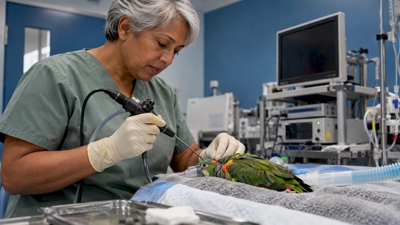
Avian air sac endoscopy (also called coelioscopy) lets a veterinarian look inside a bird's body cavity through a small incision, using the bird's own air sac system as the working space. The scope enters through a caudal air sac, usually from the left side, and the clinician can then view the lungs, air sacs, gonads, kidneys, liver and other organs without a full exploratory surgery. This guide covers why it works in birds, how access is done, what equipment suits small avian patients, and how to reduce risk. It is written for veterinarians and clinic teams.
TL;DR
- Birds have no diaphragm and no peritoneal cavity like mammals, and their air sacs are already inflated, so the scope works in a natural space with no gas insufflation.
- The usual route is a left lateral approach into the caudal thoracic air sac, then through to neighboring sacs to see the target organs.
- Small diameter rigid scopes with a good light source are the working tools. Biopsy forceps and a sample channel or accessory port extend the exam into tissue sampling.
- Anesthesia, positioning and a gentle, short procedure matter more than any single piece of equipment, because the air sacs are part of the bird's breathing mechanism.
- Image recording makes findings reviewable and helps with client communication and referral.
Table of Contents
- Why birds are suited to air sac endoscopy
- What the exam can show
- When the procedure is indicated
- Patient preparation and anesthesia
- Access: the left lateral approach
- Air sac washes and directed sampling
- Equipment for avian endoscopy
- Risks and how to lower them
- Perspective
- Getting equipped
- FAQ
- Sources
Why birds are suited to air sac endoscopy
The anatomy does most of the work. Birds have nine air sacs in total, grouped functionally into cranial and caudal systems that act as bellows for the comparatively rigid avian lung, as described in Clinical Avian Medicine. Birds also lack a diaphragm and have no peritoneal cavity analogous to that of mammals, and because the air sacs are naturally inflated, carbon dioxide insufflation is not needed to create a viewing space.

That is the key difference from mammalian laparoscopy. In a dog or cat you must distend the abdomen with gas. In a bird, the scope passes through a thin air sac wall into a space that is already open. The air sac membranes are delicate and nearly transparent, so organs lying against them can often be seen through the wall or after the scope passes into the next sac.
What the exam can show
Depending on the sacs entered and the patient's size, a skilled endoscopist can evaluate several structures during one session:
- Lungs and air sacs: surface appearance of the lung, thickening or cloudiness of sac walls, and plaques or granulomas. This is of particular interest in suspected fungal disease.
- Gonads: visual identification of testes or ovary, which is also how endoscopic sexing is done in monomorphic species.
- Kidneys and adrenal glands: position, size, color and surface contour.
- Liver, spleen and heart region: size, edge, color, and surface changes.
- Gastrointestinal organs: parts of the proventriculus, ventriculus (gizzard) and intestines may be seen from the appropriate sac.
- Syrinx and trachea: viewed through a separate route, usually through the mouth or the tracheal lumen with a flexible or very small rigid scope.
The value is that you can take targeted samples under direct vision, or decide that surgery is not warranted.
When the procedure is indicated
Clinicians typically consider endoscopy when imaging and bloodwork leave a question open about the coelomic cavity or respiratory tract. Common reasons include:
- Chronic respiratory signs, especially when Aspergillus infection is on the differential and radiographs or blood tests are not conclusive.
- Unexplained weight loss, regurgitation or lethargy where an organ biopsy would change treatment.
- Reproductive disease, including suspected ovarian or testicular pathology.
- Suspected coelomic masses or organomegaly seen on radiographs.
- Sex determination in monomorphic species.
- Collecting samples for cytology or culture from the air sacs, using direct visualization rather than blind sampling.
Matching the question to the technique is where clinical judgment comes in. Endoscopy is most useful when it will add something that other tests cannot, such as a tissue sample from a specific lesion.
Patient preparation and anesthesia
Because the air sacs are part of the respiratory system, the patient must be handled as a respiratory case, even when the primary complaint is not respiratory. Some points clinicians plan for:
- Fasting: a short fast is usually used to reduce the risk of regurgitation and to make the proventriculus and ventriculus less full. Fasting length depends on the species and size, so follow your own protocol and avoid prolonged fasting in small birds.
- Stabilization: a bird with severe dyspnea may need oxygen support and supportive care before it is anesthetized. Plan for the possibility that endoscopy is deferred.
- Anesthesia: inhalant anesthesia with an endotracheal tube is the common choice, with the ability to assist ventilation. Birds breathe by moving the keel and ribs, so positioning that restricts chest movement can compromise ventilation. The Harrison chapter linked above on avian anesthesia is a useful starting point for physiology.
- Monitoring: respiratory rate, heart rate, temperature and depth should be tracked. Small birds lose heat quickly, so a heat source is part of the setup.
- Positioning: the bird is placed in right lateral recumbency for the left lateral approach, with the upper leg drawn forward or backward to expose the flank, and the area is plucked and prepared aseptically.
Access: the left lateral approach
The approach described in the top-ranking veterinary congress proceedings is a left lateral access into the caudal thoracic air sac, then passage into the cranial thoracic air sac to see other structures. The sequence in practice looks like this:
- Choose the site. The entry point is in the flank, behind the last rib and in front of the thigh, where the body wall is thin. Landmarks vary slightly by species, so palpate before incising.
- Make a small incision. A small skin incision is made, then blunt dissection through the muscle layer with fine hemostats or scissors. Avoid vessels.
- Enter the air sac. The sac wall is penetrated with the scope, or with a sleeve and obturator, depending on your system. A change in resistance and a view of a transparent, glistening membrane tells you you are in the sac.
- Survey the sac and organs. In the caudal thoracic or abdominal sac you can usually identify the lung, kidney, gonad, adrenal and parts of the gastrointestinal tract. Move slowly, with the scope tip kept clear of the tissue, because the sac wall tears easily.
- Pass to the next sac if needed. A cranial approach through the septum is used to see the heart, liver and cranial lung. This step increases the chance of damage, so use it only when the question requires it.
- Biopsy or sample. Take samples under direct view using flexible biopsy forceps of the right size, and have a plan for bleeding.
- Withdraw and close. Remove the scope slowly and observe for bleeding. Skin closure is typically simple, and many clinicians do not suture the air sac itself.
A second approach through the ventral midline or a right-side entry is sometimes used, but the left lateral route is the standard teaching approach and the one used by most exam descriptions.
Air sac washes and directed sampling
Endoscopy is not only about looking. It also improves how you collect fluid and tissue. In a study on processing avian air sac washes, directed endoscopy was used to obtain the wash samples, and the authors compared handling factors that might affect preservation of cell morphology. The takeaway for the clinic is practical: when you can see where the lesion is, you can sample it directly, and the quality of what the lab receives depends on how you handle and preserve the fluid.
Compared with blind sampling:
| Approach | What you gain | What you give up |
|---|---|---|
| Endoscopy with direct sampling | Sample from a visible lesion, biopsy under view, images for the record | Needs equipment, anesthesia and training |
| Air sac wash without visualization | Faster and simpler | Less certainty about where the sample came from |
| Radiographs and bloodwork alone | Non-invasive, quick | Often non-specific for air sac and lung disease |
Most cases use these methods together. Endoscopy is typically reserved for when the other tools have not answered the question.
Equipment for avian endoscopy
Bird patients are small, so the instrument must be small too. The same logic applies to reptiles, where a 2.7 mm scope is a common starting point, and the benefits of narrow instruments are covered in our guide to small diameter endoscopes in veterinary practice.
A typical avian setup includes:
- A small diameter rigid endoscope for the coelomic exam, often with a sheath for irrigation or instrument passage. Choose a diameter that suits the smallest species you see.
- A cold light source with enough output for a clean image. Light that is too weak gives a dark, grainy picture; too strong a source can warm the tip, so keep tip contact with tissue minimal.
- A camera or videoscope system with recording, so findings can be saved to the record. Our guide to optimizing endoscope video recording covers settings and workflow.
- Biopsy forceps sized to the sheath, as well as grasping forceps for foreign material.
- Flexible scopes for the trachea and syrinx, where the rigid instrument does not fit.
- Sterilization supplies and a reprocessing routine that protects delicate optics. Our common endoscopic procedures guide and small animal endoscopy tips cover general handling that applies here.
For clinics that want to see the options, our rigid scopes, arthroscopes and otoscopes category and flexible biopsy forceps are the relevant pages.
Risks and how to lower them
Air sac endoscopy is considered a minimally invasive technique, but it is not risk free. The main concerns are:
- Respiratory compromise. Anesthesia in birds with air sac or lung disease needs particular care. Have oxygen and assisted ventilation ready, and keep the procedure short.
- Hemorrhage. Biopsy of the liver, kidney or gonad can bleed. Choose the site carefully, use the smallest effective forceps, and watch the site before withdrawing.
- Air sac or organ damage. Sudden movement or a scope pushed against tissue can tear the sac or injure an organ. Hold the scope steadily and move with the image, not ahead of it.
- Fluid or fungal contamination. If the air sac contains fluid or plaques, avoid spreading material to other sacs. Sample from the most affected area first when possible.
- Hypothermia. Small birds lose heat quickly during plucking, prepping and anesthesia.
- Infection. Use aseptic technique and properly reprocessed instruments, because a contaminated scope can seed the air sacs.
Case selection also matters. A critically ill bird may be too unstable for anesthesia, and the decision to proceed rests with the attending veterinarian.
Perspective
Small birds are one of the clearest cases for choosing the right-sized scope. The patient leaves very little margin, so the image has to be good, the instrument has to be slim, and the setup has to be quick to deploy. Clinics often hesitate because they assume avian endoscopy requires a hospital-grade human system. We built our offering around the opposite idea: portable, affordable systems with video recording, so a practice can do the exam in-house and keep a record of what it saw.
We also believe in support. Whether a clinic is buying its first scope or adding a smaller one for exotics, it should be able to ask questions about sheath size, light output, forceps compatibility and care, and get answers on the same day. That is why we offer 24/7 support and rapid shipping, and why we repair endoscopes too.
Our advice is to start with the questions you ask most often. If your caseload is mostly small birds and reptiles, begin with a slim rigid scope and a reliable light source. Add flexible instruments for the airway later.
Endoscope
Getting equipped
If you are setting up or expanding an exotics endoscopy service, our team can help you match scope diameter, light source and accessories to your species mix. Browse the veterinary endoscopy equipment page, or look at our medical endoscopes and accessories for light sources, valves, pumps and cleaning tools.
If an instrument needs service, we also handle endoscope repairs, so a damaged scope does not leave the procedure room empty for long. Visit 1800endoscope.com to start.
FAQ
Do birds need CO2 insufflation for endoscopy?
No. Birds have naturally inflated air sacs and no diaphragm or peritoneal cavity like mammals, so the scope works in an existing space. Veterinary Practice News describes this anatomical advantage. Gas insufflation is therefore not part of the standard technique.
Which air sac is usually entered first?
The common teaching approach is a left lateral entry into the caudal thoracic air sac. From there the scope may be passed into the cranial thoracic sac to see additional organs. Your choice depends on the target organ and the size of the bird.
Can air sac endoscopy help diagnose fungal disease?
It can help by letting the clinician see plaques or granulomas and collect samples from them directly. Aspergillus infection is a well-described problem in birds, and the final diagnosis usually combines visual findings with cytology, culture, histopathology or other tests.
What size scope is used for small birds?
Small diameter rigid scopes are used, with the exact size chosen for the species and patient. A narrow instrument lowers the risk of tissue trauma and is easier to maneuver within the sac. See our small diameter endoscope guide for more detail.
Is the procedure safe for sick birds?
It carries real anesthetic risk, especially in birds with respiratory disease. Stabilization, oxygen support, careful monitoring and a short procedure are part of safe practice, and the veterinarian decides whether a bird is fit for the exam.
Sources
- Clinical Avian Medicine: avian anesthesia chapter
- Avian Anatomy Aids Endoscopic Procedures, Veterinary Practice News
- Aspergillus infections in birds: a review, Avian Pathology