
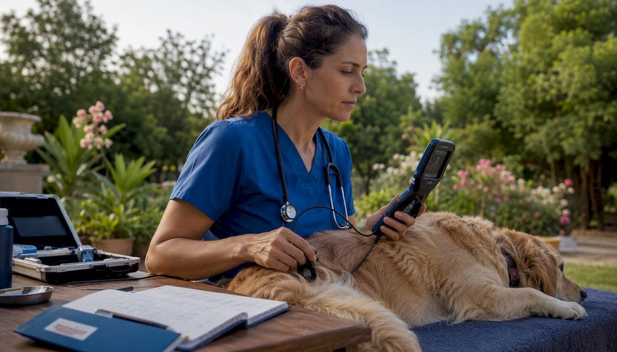
Video imaging is a frontline diagnostic and documentation tool in modern veterinary practice. It delivers real-time, noninvasive visualization, creates a permanent medical record, and increasingly feeds AI-assisted analysis workflows. Three practical takeaways before you read further: endoscopy and cine ultrasound are the most clinically accessible video modalities in daily practice; acquisition quality and standard views matter more than equipment brand; and AI tools show genuine promise but require species-specific validation before you trust them clinically.
A systematic review of deep learning in veterinary diagnostics screened 422 publications and identified 39 primary research studies, with radiography and cytology each representing a large portion of included work and photo/video imaging accounting for a small fraction. That gap tells you where the field is and where it is heading. A 2026 modified-Delphi study confirmed that several physical exam components can be performed via video, while 15 cannot, which sets a realistic ceiling for telemedicine-based video assessment. The American College of Veterinary Radiology (ACVR) and the Merck Veterinary Manual emphasize that imaging quality and standard views are non-negotiable for defensible diagnosis.
Table of Contents
- What is the role of video imaging in veterinary diagnostics?
- Clinical applications of video imaging across species
- How do you select and operate video imaging equipment?
- Where does AI fit in veterinary video and image analysis?
- Why acquisition quality determines diagnostic value
- How do you integrate video imaging into clinic workflow?
- What are the real limitations and pitfalls of video imaging?
- What video imaging trends should veterinarians watch?
- Key Takeaways
- The case for staged adoption, not wholesale commitment
- 1800endoscope carries the scopes your clinic actually needs
- Useful sources for further reading
What is the role of video imaging in veterinary diagnostics?
Video imaging sits within a broader toolkit that includes radiography, ultrasonography, computed tomography (CT), magnetic resonance imaging (MRI), nuclear medicine, and endoscopy-based video modalities. Each has a distinct clinical niche, and understanding where video-based techniques fit helps you triage cases efficiently.
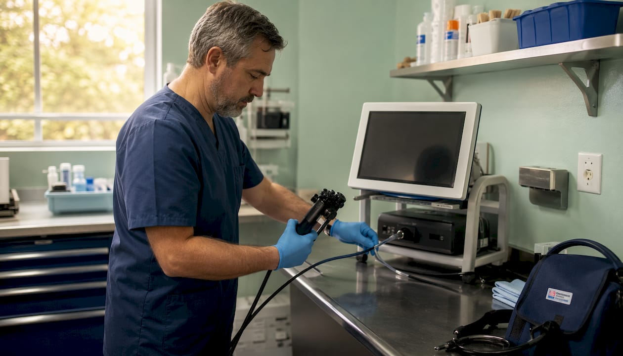
Radiography and ultrasonography remain the workhorses of veterinary practice, driven by cost, availability, and the absence of anesthesia requirements in most cases. CT and MRI offer superior anatomic detail and soft-tissue contrast, but both carry higher cost, anesthesia time, and logistical overhead that limit routine use. MRI is the preferred modality for brain parenchyma evaluation and chronic equine foot lameness. Nuclear medicine remains largely referral-center territory.
Video-based modalities occupy a distinct space: they provide dynamic, real-time visualization of luminal structures, moving anatomy, and surgical fields that static images simply cannot capture. The table below maps each major modality to its primary indications and key trade-offs.
| Modality | Best For | Key Advantages | Key Limitations |
|---|---|---|---|
| Radiography | Skeletal, thoracic, abdominal survey | Fast, low cost, widely available | No real-time motion; limited soft-tissue contrast |
| Ultrasonography (incl. cine/Doppler) | Soft tissue, cardiac, reproductive, GI | Real-time, portable, no anesthesia | Operator-dependent; limited through bone/gas |
| CT | Complex fractures, thoracic/abdominal detail, oncology staging | High-resolution 3D reconstruction | Anesthesia required; high cost; limited portability |
| MRI | CNS, soft-tissue orthopedics, equine foot | Superior soft-tissue contrast | Long scan time; anesthesia; specialist access |
| Endoscopy/Videoscopy | Airway, GI, urinary, reproductive luminal structures | Direct visualization, biopsy capability, video record | Invasive access; sedation often needed |
| Fluoroscopy | Dynamic swallowing, orthopedic reduction, contrast studies | Real-time motion capture | Radiation; specialist equipment |
| Video-assisted laparoscopy/arthroscopy | Abdominal organs, joints | Minimally invasive surgery and diagnosis | Requires training; general anesthesia |
Species access and economics shape these choices significantly. In equine practice, field portability drives most decisions: ultrasound and flexible endoscopy travel to the patient. In small-animal practice, the full modality range is more accessible, but CT and MRI still require referral in most general practices. Production animal medicine leans heavily on portable ultrasound and basic endoscopy because per-animal economics rarely justify advanced imaging.
Clinical applications of video imaging across species
The real value of video imaging technology for animals becomes clear when you look at specific clinical scenarios. Across species and body systems, video-based modalities change what you can see, what you can do, and how you document it.
Respiratory and airway
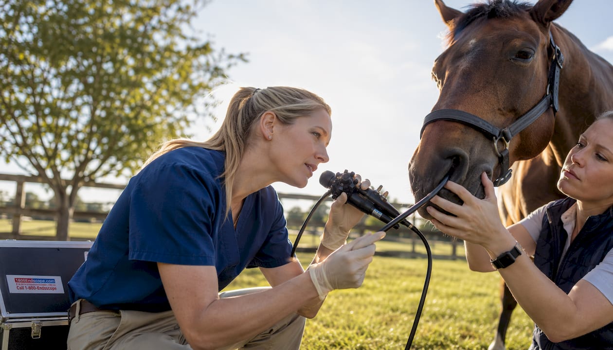
Flexible endoscopy is the standard of care for upper airway evaluation in horses. Dynamic respiratory endoscopy performed on a treadmill or during ridden exercise captures intermittent obstructions, such as recurrent laryngeal neuropathy or dorsal displacement of the soft palate, that a resting scope simply misses. In small animals, video laryngoscopy allows direct visualization of laryngeal function during light anesthesia, which is the definitive approach for laryngeal paralysis diagnosis. Bronchoscopy extends the view to the lower airways for bronchoalveolar lavage, foreign body retrieval, and mass biopsy.
Gastrointestinal
Gastroscopy is the primary diagnostic tool for equine gastric ulcer syndrome (EGUS) grading. In small animals, upper GI endoscopy evaluates chronic vomiting, protein-losing enteropathy, and esophageal disease with biopsy capability that no other noninvasive method matches. Colonoscopy adds visualization of the large bowel for mass lesions, inflammatory disease, and foreign material. A 3-meter economy gastroscope covers most small-animal and equine gastric work without the cost of a full endoscopy tower.
Urinary and reproductive
Cystoscopy in dogs and cats allows direct visualization of the bladder mucosa, ureteral openings, and urethra, which is particularly useful for ectopic ureter diagnosis and transitional cell carcinoma staging. In mares, video-assisted uterine endoscopy evaluates endometrial health and guides biopsy placement far more precisely than blind techniques.
Musculoskeletal and intraoperative
Arthroscopy in horses and dogs provides both diagnostic and therapeutic access to joints with minimal morbidity compared with open arthrotomy. Laparoscopy in small animals and horses allows organ biopsy, cryptorchid castration, and ovariectomy under video guidance. Fluoroscopy guides fracture reduction and implant placement in real time.
Three clinical vignettes
Companion animal, chronic cough: A 7-year-old Labrador presents with a six-month history of productive cough unresponsive to antibiotics. Thoracic radiographs show a mild bronchial pattern but no mass. Bronchoscopy reveals a partially obstructing endobronchial mass at the right caudal bronchus. Biopsy confirms primary lung carcinoma. Without video imaging, the diagnosis would have required CT or open thoracotomy.
Equine recurrent colic: A 12-year-old Warmblood with recurrent right dorsal displacement episodes undergoes gastroscopy, which reveals grade 3 EGUS. Dietary management and omeprazole therapy resolve the colic episodes over 60 days. The video record documents ulcer grade at baseline and at recheck, supporting treatment decisions and client communication.
Production animal, reproductive screening: A dairy herd with declining conception rates undergoes video-assisted uterine endoscopy on a sample of repeat-breeder cows. Endometritis is confirmed in 40% of examined animals, guiding targeted intrauterine therapy and improving herd reproductive efficiency.
Pro Tip: For chronic cough, choose bronchoscopy over radiography alone when the radiograph is equivocal or the cough is productive. For recurrent colic in horses, gastroscopy before initiating empirical ulcer therapy gives you a grade to treat to, not just a suspicion to manage.
How do you select and operate video imaging equipment?
Equipment decisions come down to four variables: the species you treat, the body systems you image most, your budget, and your clinic's physical setup. Getting the spec wrong costs you diagnostic yield; getting it right means the scope earns its keep on day one.
Equipment checklist
- Insertion tube diameter: 2.8–4 mm for feline/small-dog urinary and nasal work; 8–10 mm for equine airway and gastric; 10–13 mm for large-animal gastroscopy
- Working length: 1 m for small-animal upper GI; 3 m for equine gastroscopy; 60–90 cm for arthroscopy/laparoscopy
- Camera resolution: 1080p HD minimum for diagnostic-quality capture; 4K where budget allows
- Light source: LED preferred for portability and consistent color temperature; xenon for highest brightness in large-lumen work
- Monitor and recorder: integrated monitor with SD card or USB recording simplifies documentation; HDMI output to external monitor for surgical use
- Connectivity: wireless or USB output for EMR/PACS integration and telemedicine sharing
- Sterilization compatibility: confirm high-level disinfection (HLD) or autoclave compatibility per manufacturer spec before purchase
Procurement reference
| Scope Type | Diameter | Working Length | Sterilization | Typical Use |
|---|---|---|---|---|
| Small-animal flexible videoscope | 2.8–5 mm | 60 cm | HLD (glutaraldehyde/OPA) | Nasal, urinary, upper GI |
| Equine airway videoscope | 8–10 mm | — | HLD | Upper airway, guttural pouch |
| Equine gastroscope | 10–13 mm | 3 m | HLD | Gastric ulcer evaluation |
| Rigid arthroscope | 2.8–4 mm | 10 cm | Autoclave | Joints, laparoscopy |
| Portable field videoscope | 6 mm | — | HLD | Airway, dental, field use |
The portable 6mm airway videoscope with direct monitor and SD card recording is a practical starting point for mixed practices that need field capability without a full tower. For equine-specific airway work, an 8mm USB field scope with SD card recording covers treadmill and stall-side exams.
Reprocessing is where most clinical errors occur. After each procedure: pre-clean at bedside, leak-test before immersion, perform manual cleaning with enzymatic detergent, then complete HLD per the manufacturer's validated contact time and concentration. Never skip the leak test; a single immersion of a damaged scope can destroy the insertion tube. Store scopes hanging vertically or in a dedicated case, never coiled tightly.
Warranty and service considerations matter as much as purchase price. Confirm that the manufacturer offers U.S.-based repair service and that loaner scopes are available during servicing. A scope out of service for six weeks is a significant revenue gap in a busy practice. For troubleshooting common issues, the videoscope troubleshooting guide covers the most frequent failure modes and their fixes.
Pro Tip: Portability and image quality are not mutually exclusive anymore. A 1080p portable scope with an integrated monitor and SD card recording gives you diagnostic-grade video in the field. The trade-off is working channel diameter: portable scopes often have a 2.0 mm or no working channel, which limits biopsy capability. Know before you buy whether you need biopsy access.
Where does AI fit in veterinary video and image analysis?
AI and deep learning are genuinely changing what is possible in veterinary imaging, but the evidence base is uneven and the gap between published accuracy and clinical reliability is wider than most vendor materials suggest.
The systematic review of 39 primary deep learning studies in veterinary diagnostics found that radiography and cytology dominate current AI work, with photo and video imaging representing only about 5% of included studies. Within that small video-specific body of work, results are promising: convolutional neural network (CNN) models using MobileNetV2, InceptionV3, and VGG variants achieved high accuracy on ophthalmic image classification tasks using smartphone-derived images. That is a meaningful result, but it applies to a narrow, well-annotated task, not to general video diagnostics.
The more ambitious direction is behavioral phenotyping and pose estimation. The AnimalFormer framework demonstrated how combining GroundingDINO, HQ-SAM, and ViTPose on sheep video datasets can extract activity patterns, grazing behavior, and postural analytics without physical markers. Reliable pose estimation requires multi-camera setups; single-camera models are prone to occlusion errors and misclassification when the recording geometry differs from training data.
The central validation challenge in veterinary video AI is not model architecture. It is dataset quality. Species-specific physiological variation means that a model trained on human colonoscopy video will not transfer to canine GI endoscopy without retraining on annotated veterinary data. The same applies across species: a bovine lameness model trained on Holstein gait data may perform poorly on Angus or dairy goat subjects. Before adopting any AI tool for clinical use, ask the vendor for external validation data on your target species, the dataset size and diversity, and the sensitivity and specificity on a held-out test set.
Clinician validation checklist for AI imaging tools
When evaluating any AI-assisted video or image analysis tool, work through these questions before clinical deployment:
- Sample size and diversity: Was the training dataset large enough and diverse enough to cover your patient population's breed, age, and body condition variation?
- Sensitivity and specificity: Are both reported? A tool with 95% sensitivity but 60% specificity generates too many false positives for routine screening.
- External validation: Was the model tested on data from a different institution than the one that trained it? Internal validation inflates performance estimates.
- Species specificity: Was the model trained and validated on your target species, not a proxy population?
- Explainability: Does the tool provide a visual explanation (e.g., Grad-CAM heatmap) of what drove its output? Black-box outputs are harder to defend clinically.
- Workflow integration: Can the tool ingest your existing video file formats and output results in a format your EMR can store?
Why acquisition quality determines diagnostic value
The best scope in the world produces useless data if the image is blurry, the angle is wrong, or the clip is too short to review. Acquisition quality is the single highest-leverage variable in video imaging, and it is entirely within your control.
The Merck Veterinary Manual's radiography guidance makes the point clearly for static imaging: two orthogonal views are standard practice for musculoskeletal cases, and missing a standard view can mean a missed lesion and a medicolegal exposure. The same principle applies to video: a single-pass endoscopic clip that misses the fundus or the carina is an incomplete examination, regardless of how good the scope is.
Acquisition best practices
- Steady frames: advance the scope slowly; rapid movement creates motion blur that obscures mucosal detail
- Two orthogonal views for radiographs: lateral and ventrodorsal/dorsoventral as minimum standard
- Lighting and angulation for endoscopy: keep the tip 5–10 mm from the mucosa for optimal illumination; adjust insufflation to open luminal structures before capturing
- Cine capture settings: record at minimum 30 fps for smooth playback; 60 fps for fast-moving structures (cardiac, laryngeal)
- Clip length: capture at least 30 seconds of continuous video at each anatomical landmark; short clips miss intermittent findings
- White balance: calibrate before each procedure to ensure accurate mucosal color representation
Documentation metadata
Every video clip and image should carry the following metadata, either embedded in the file or recorded in the patient record:
- Patient ID and species/breed
- Date, time, and operator name
- Modality and scope ID (serial number)
- Sedation or anesthesia status and agent
- Relevant vitals at time of imaging (heart rate, SpO2 if applicable)
- Anatomical location and view orientation
Imaging is a permanent medical document. A poorly acquired clip that leads to a missed diagnosis is not just a clinical failure; it is a liability. Courts and licensing boards treat imaging records the same way they treat surgical notes. Optimizing endoscope video recording from the start of a procedure, not as an afterthought, is the professional standard.
Pro Tip: Adopt a consistent file-naming convention from day one: PatientID_Date_Modality_Operator (e.g., 00123_20260315_GastroScope_JSmith). It takes 10 seconds per procedure and saves hours when you need to retrieve a clip for a referral, recheck, or legal review.
How do you integrate video imaging into clinic workflow?
Adding video imaging to a practice is not just an equipment purchase. It changes triage logic, staffing requirements, data storage needs, and client communication. A staged approach prevents the common failure mode: buying a scope that sits in a cabinet because the workflow was never built around it.
Workflow integration
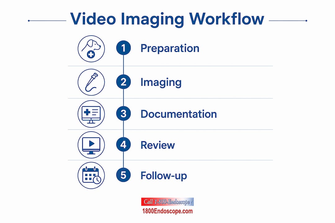
Triage: Video imaging should enter the decision tree at the point where static imaging is equivocal or insufficient. A chronic cough case that has had two sets of thoracic radiographs without a diagnosis is a bronchoscopy candidate, not a third radiograph candidate. Build this decision point into your triage protocol explicitly.
Imaging and documentation: Assign one staff member per procedure as the dedicated recorder. Their job is to confirm the scope ID is logged, start recording before scope insertion, annotate landmarks verbally on the audio track, and confirm the clip is saved before the scope is withdrawn. This takes two minutes and prevents the most common documentation failure.
Review and follow-up: Video clips should be reviewed at full length before the case is closed, not just during the procedure. Findings missed in real time are often visible on review. Schedule a 10-minute post-procedure review as standard. For complex cases, remote visual inspection and telemedicine consultation allow a specialist to review the clip without the patient traveling.
Training and credentialing
Endoscopy and video-assisted procedures require supervised training before independent practice. The number of supervised procedures needed varies by modality: upper GI endoscopy in small animals is generally achievable in 20–30 supervised cases; equine gastroscopy requires fewer cases but more physical technique. Continuing education through the American College of Veterinary Internal Medicine (ACVIM) and specialty residency programs provides structured pathways. Technicians can be trained to assist with scope preparation, patient positioning, and documentation, which frees the clinician to focus on the procedure itself.
Data governance and consent
U.S. veterinary practices are not subject to HIPAA, which applies to human health information, but client data and patient records carry state-level privacy obligations and professional licensing board requirements. Key points:
- Obtain written informed consent before recording video for telemedicine sharing or educational use
- Store video files on encrypted, access-controlled servers or PACS systems
- Retain imaging records per your state veterinary board's minimum retention period (typically 3–5 years for adult animals)
- When sharing clips for specialist consultation, use secure file transfer, not consumer email or messaging apps
The 2026 Delphi consensus on virtual exams confirms that video is supportive, not a replacement for hands-on examination components. Document which components were performed in person and which were assessed via video in every telemedicine encounter. This protects you clinically and legally.
Pro Tip: Partner with a U.S. veterinary clinic that already has a telemedicine workflow, such as Animal Care of Hobe Sound, to benchmark your consent forms and data-sharing protocols against a working model before you build your own from scratch.
What are the real limitations and pitfalls of video imaging?
Video imaging is not a universal solution, and overconfidence in the technology causes real diagnostic errors. Knowing where the method fails is as important as knowing where it succeeds.
Common pitfalls
- Poor patient positioning or inadequate sedation: Movement artifact and suboptimal angles are the most common causes of non-diagnostic studies. Inadequate sedation in an endoscopy case produces a dangerous and useless procedure.
- Low-resolution capture: Recording at compressed resolution or low frame rate degrades mucosal detail. A 480p clip from a 1080p scope is a documentation failure, not an equipment failure.
- Misapplied AI models: Using a model validated on one species or breed for a different population without revalidation is a known source of error. This applies to both image classification and behavioral phenotyping tools.
- Incomplete documentation: Missing scope ID, operator name, or sedation status in the record creates gaps that are difficult to defend in a complaint or litigation.
- Overreliance on video alone: Video endoscopy shows the mucosal surface. It does not replace histopathology, culture, or cross-sectional imaging for submucosal or extraluminal disease.
- Insufficient training: Attempting advanced procedures (e.g., equine tracheal wash via bronchoscope, laparoscopic biopsy) without adequate supervised experience increases complication risk and reduces diagnostic yield.
Mitigation strategies
A QA checklist completed before every procedure covers the highest-risk failure points: patient ID confirmed, scope leak-tested, recording started, sedation documented. Second reads on video clips by a second clinician or a specialist via telemedicine catch findings missed in real time. Standardized protocols for each procedure type (scope diameter, insufflation pressure, clip length, landmark documentation) reduce variability across operators.
When to escalate: refer to a specialty imaging center when CT or MRI is needed for CNS disease, complex orthopedic staging, or oncology workup; when a procedure requires general anesthesia and your facility lacks monitoring capability; or when a video finding is ambiguous and the clinical stakes are high. Advanced imaging methods for veterinarians at referral centers often include interventional radiology and fluoroscopy-guided procedures that general practices cannot replicate.
Economic constraints are real. CT and MRI require capital investment, specialist staffing, and anesthesia infrastructure that most general practices cannot justify. The practical answer is a tiered approach: use ultrasound and endoscopy as first-line video modalities, build referral relationships with imaging centers for CT/MRI cases, and reserve advanced imaging for cases where the clinical decision genuinely depends on it.
What video imaging trends should veterinarians watch?
The next three to five years will bring meaningful changes to how video imaging technology for animals is deployed, analyzed, and integrated into clinical workflows. These are the trends worth tracking now.
- Multimodal AI integration: Models that fuse video, static images, and clinical metadata are moving from research to early commercial deployment. The main barrier remains species-specific physiological variation; human-derived models rarely transfer directly to veterinary populations without retraining.
- Pose estimation and behavioral phenotyping: Livestock and companion animal behavioral analytics from video are advancing rapidly. Reliable outputs require multi-camera setups and high-quality annotated datasets; single-camera systems remain prone to occlusion errors.
- Portable HD and wireless scopes: 1080p and 4K portable videoscopes with wireless streaming are now available at price points accessible to general practice. This changes field diagnostics for equine and production animal practitioners significantly.
- Cloud-based tele-endoscopy: Secure cloud platforms for real-time or asynchronous specialist review of endoscopy video are entering the U.S. market. This extends specialist access to rural and mixed practices without patient transport.
- Improved 3D/2D fusion imaging: Combining endoscopic video with CT or ultrasound data for intraoperative navigation is an active research area, primarily in academic centers, but will reach referral practice within five years.
- Augmented intraoperative guidance: Overlay of anatomical landmarks and tissue identification on live laparoscopy or arthroscopy video is in early clinical validation. Expect commercial systems in equine and small-animal surgery within the mid-term horizon.
For procurement planning: the portability and wireless connectivity trend means that a scope purchased today should have USB or Wi-Fi output as a baseline spec, not an optional upgrade. For training planning: pose estimation and behavioral AI tools will require staff who understand multi-camera setup and video annotation, not just scope operation.
Key Takeaways
Video imaging is most valuable in veterinary practice when acquisition quality is prioritized, modalities are matched to clinical indications, and AI tools are validated on species-specific datasets before clinical use.
| Point | Details |
|---|---|
| Video imaging as frontline tool | Endoscopy and cine ultrasound provide real-time, noninvasive visualization that static imaging cannot replicate. |
| Acquisition quality is highest priority | Poor positioning, low frame rate, or missing landmarks produce non-diagnostic studies regardless of equipment quality. |
| AI requires species-specific validation | A systematic review found only 39 primary DL studies in veterinary diagnostics; video-specific evidence accounts for about 5% of included studies. |
| Equipment selection by species and system | Match scope diameter, working length, and sterilization method to your most common procedures before purchasing. |
| 1800endoscope for clinical procurement | 1800endoscope offers portable videoscopes, equine airway scopes, gastroscopes, and rigid endoscopy systems suited to the clinical uses described in this guide. |
The case for staged adoption, not wholesale commitment
The most common mistake practices make with video imaging is treating it as an all-or-nothing investment. A full endoscopy tower with a gastroscope, bronchoscope, and colonoscope is the right answer for a high-volume internal medicine practice. For a mixed or general practice, it is often the wrong starting point.
Start with one scope that covers your highest-volume indication. For most small-animal practices, that is a flexible upper GI scope in the 8–10 mm range. For equine practices, it is an airway scope. Use it for 90 days, track how many cases it changes, and measure the time from presentation to diagnosis. Those numbers tell you whether to expand.
The documentation workflow matters as much as the equipment. Practices that add a scope without building a recording and storage protocol end up with a library of unlabeled clips that cannot be retrieved, reviewed, or shared. That is not a technology problem; it is a workflow problem. Solve it before the scope arrives, not after.
AI tools deserve cautious optimism. The research is real and the trajectory is clear, but the gap between published accuracy on curated datasets and reliable performance in a busy general practice is still wide. Evaluate any AI imaging tool the same way you would evaluate a new diagnostic test: ask for sensitivity, specificity, and external validation data on your target species. If the vendor cannot provide those numbers, the tool is not ready for clinical use.
1800endoscope carries the scopes your clinic actually needs
Clinicians who have worked through the modality and equipment guidance in this article often arrive at the same question: where do you source a reliable, affordable scope without a six-month procurement cycle?

1800endoscope stocks portable videoscopes, equine airway and dental inspection scopes, economy gastroscopes, and a full rigid endoscopy catalog for arthroscopy and laparoscopy applications. Every system ships with HD video recording capability, and most portable units include an integrated monitor and SD card storage so you can document from the first procedure. The full catalog covers light sources, biopsy forceps, valves, and cleaning accessories alongside the scopes themselves. Lead times are short, and the product pages include spec sheets so your procurement team can confirm sterilization compatibility and working channel dimensions before ordering. Browse the catalog or contact 1800endoscope directly to match a scope to your clinical workflow.
Useful sources for further reading
| Source | Why It Matters |
|---|---|
| Deep learning in veterinary diagnostics: systematic review (Frontiers in Veterinary Science) | Screened 422 publications; 39 primary DL studies; quantifies the current evidence base and identifies that photo/video imaging accounts for a small fraction (about 5%) of included studies. |
| CT, MRI, and nuclear medicine in veterinary medicine (PMC) | Covers advanced modality indications, anesthesia requirements, and cost/access constraints; essential context for triage decisions. |
| Ultrasound and imaging modalities in veterinary practice (PMC) | Reviews ultrasound (including Doppler and 3D) as a cost-effective, real-time option for soft-tissue and reproductive indications across species. |
| Virtual companion animal physical exams: Delphi consensus | Identifies 19 exam components feasible via video and 15 that are not; sets realistic expectations for telemedicine-based video assessment. |
| AnimalFormer: multimodal vision framework for livestock (arXiv) | Demonstrates GroundingDINO, HQ-SAM, and ViTPose integration for behavioral phenotyping from sheep video; key reference for AI and pose estimation trends. |
| Radiography of animals, Merck Veterinary Manual | Standard reference for acquisition quality, positioning, and the medicolegal importance of complete imaging records. |
| 3D-AI BehaviorAtlas, TSE Systems | Illustrates multi-camera pose estimation requirements and the limitations of single-camera behavioral AI models. |