
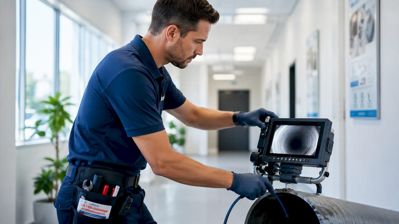
Inspection camera zoom functions are the core features that determine how accurately a technician can magnify, examine, and diagnose a target area without physical access. The two primary zoom types are optical zoom and digital zoom, and understanding the difference between them is not optional for professionals. Optical zoom physically adjusts the lens focal length to produce a magnified image with no loss of resolution. Digital zoom enlarges the existing pixel data through software interpolation, which degrades image clarity. For industrial NDT inspections and veterinary diagnostics alike, optical zoom preserves resolution in ways that digital zoom simply cannot match. Getting this distinction right is the foundation of every effective inspection workflow.
What are the different types of zoom functions in inspection cameras?
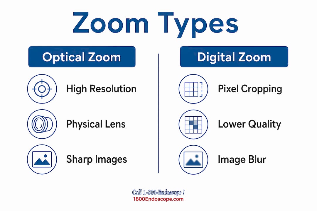
Optical zoom and digital zoom operate on entirely different principles, and the gap in performance matters enormously in the field.
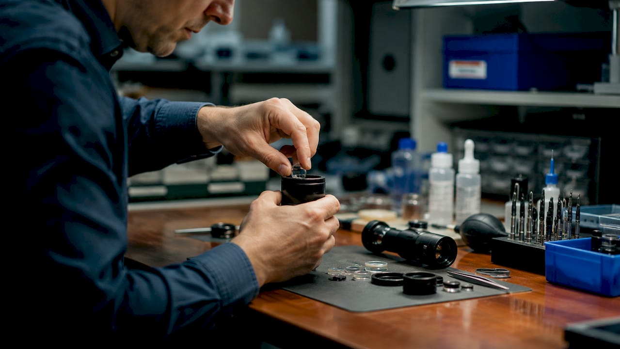
Optical zoom works by physically moving lens elements to change the focal length of the camera. The image sensor receives a genuinely magnified image, so resolution stays constant regardless of the zoom level. The Sony FCB-ER9500, a widely referenced industrial inspection module, delivers 25x lossless optical zoom with a focal length range of 5.1mm to 127.5mm. That range lets a single camera function as both a close-up microscope and a distant surveying tool without switching equipment.
Digital zoom works differently. The camera crops the center of the image and enlarges it using pixel interpolation. No new visual data is added. The result is a larger image with the same or less actual detail than the original. For reviewing general site conditions, digital zoom can be convenient. For detecting a hairline crack in a weld or a mucosal lesion in a veterinary airway exam, it is not reliable.
Pipeline inspection cameras illustrate how both zoom types are used together in practice. A typical field configuration combines 20–40x optical zoom with 10–15x digital zoom, giving operators a wide magnification range for rapid, non-invasive condition assessments. The optical zoom handles the critical diagnostic work, while digital zoom assists with general orientation and documentation.
- Optical zoom: Adjusts focal length physically; resolution is preserved at all magnification levels.
- Digital zoom: Crops and interpolates pixels; convenient but degrades image fidelity.
- Hybrid configurations: Combine both types; optical zoom handles precision work, digital zoom handles general review.
- Zoom ratios: Industrial cameras commonly offer 20–40x optical zoom; veterinary scopes typically use lower ratios suited to confined anatomical spaces.
Pro Tip: Always confirm whether a camera's stated zoom ratio refers to optical or digital zoom before purchasing. A "40x zoom" label that refers to combined optical and digital zoom is not the same as 40x optical zoom.
How does zoom level impact inspection accuracy?
Zoom level directly determines whether a technician can detect a defect or miss it entirely. This is not a matter of image preference. It is a matter of diagnostic validity.
Optical zoom is mandatory for any inspection task that involves measurement or AI-assisted defect recognition. When a camera uses optical zoom, the pixel data represents real, captured detail. An AI defect recognition algorithm fed optically zoomed imagery has accurate data to work with. Feed it digitally zoomed imagery and the algorithm is working from interpolated guesses, not real surface data. The difference shows up in false negatives and missed defects.
Microscopic defects are the clearest test of zoom quality. Weld porosity, hairline cracks in metal structures, and early-stage corrosion pitting are all features that measure in fractions of a millimeter. At low magnification, they are invisible. At high optical zoom, they are clearly defined. Veterinary applications face the same challenge: identifying early mucosal inflammation, foreign body fragments, or dental pathology in a horse's oral cavity requires genuine magnification, not a pixelated approximation.
"Zoom facilitates faster and more accurate diagnostics, reducing labor costs and equipment downtime in inspection workflows. The ability to identify defects remotely at high magnification eliminates the need for repeated physical access to confined or hazardous areas."
HD sensors combined with optical zoom create a compounding advantage. A 4K sensor at 25x optical zoom produces images where fine surface details are both magnified and sharp. That combination allows a single inspection pass to capture data that would otherwise require multiple visits or destructive testing. The result is a direct reduction in labor hours and equipment downtime, which translates to measurable cost savings for inspection firms and veterinary practices alike.
Digital zoom's limitations become most visible in critical diagnostic tasks. Operators who rely on digital zoom for crack detection risk producing inspection reports based on interpolated image data. That is a liability issue, not just a technical one. Any inspection report used to certify structural integrity or support a veterinary diagnosis must be based on real image data.
What are the challenges and best practices when using zoom functions?
High zoom magnification amplifies every problem in the imaging chain, not just the defect you are looking for. Lighting, camera stability, and movement speed all become critical variables at high zoom levels.
Managing lighting at high zoom
Lighting is the most commonly mismanaged variable in zoomed inspections. Excessive LED brightness in wet or confined spaces washes out surface detail, making zoom ineffective regardless of the optical quality of the lens. The correct approach is to start with minimal lighting and increase it gradually until detail becomes visible under zoom. In sewer and pipeline inspections, reflective pipe walls amplify this problem significantly. In veterinary endoscopy, mucosal surfaces can similarly reflect light in ways that obscure the very features you are trying to examine.
Maintaining camera stability under zoom
Camera movement that is barely noticeable at 1x magnification becomes severe image blur at 20x. Stability is not optional at high zoom levels.
- Move the camera at approximately 1 foot per second inside pipes or confined spaces. Faster movement at high zoom produces motion blur that makes footage unusable for defect documentation.
- After each turn or directional change, pause and allow the camera's internal stabilization or pendulum leveling mechanism to settle before zooming in on a target area.
- Use articulating camera heads where available. A four-way articulating tip gives you directional control without physically rotating the insertion tube, which reduces vibration transmitted to the image sensor.
- Brace the insertion tube against the pipe wall or anatomical structure when holding a zoomed position for documentation. Even minor hand tremor becomes visible at high magnification.
- Review footage at full resolution before concluding an inspection. Compression artifacts in recorded video can mimic surface defects at high zoom, so always verify findings against the original uncompressed feed where possible.
Pro Tip: When inspecting welds or veterinary tissue at maximum optical zoom, lock the camera position and let the image stabilize for two to three seconds before capturing a still image. Moving and shooting simultaneously at high zoom almost always produces a blurred result.
Avoiding digital zoom in critical tasks
Digital zoom compromises data integrity in any inspection where the findings will be used for certification, reporting, or clinical diagnosis. Reserve digital zoom for general orientation and documentation of large-scale features. Switch to optical zoom the moment you need to confirm a specific defect or anatomical finding.
Cleaning and maintenance
Zoom lens elements and camera heads accumulate debris, moisture, and biological material in field conditions. A contaminated lens at high zoom magnifies the contamination along with the target. Clean the camera tip before each inspection using the manufacturer-recommended method. For veterinary scopes, follow disinfection protocols appropriate to the procedure type. A clean lens at 10x optical zoom produces better diagnostic images than a contaminated lens at 25x.
How to use inspection camera zoom functions effectively in the field
Translating zoom knowledge into a repeatable field workflow separates technicians who get reliable results from those who struggle with inconsistent image quality. The workflow below applies to both industrial and veterinary inspection scenarios.
Understanding how inspection cameras transform diagnostics in both fields starts with a consistent operating sequence.
- Set baseline magnification first. Begin every inspection at the lowest useful zoom level to orient yourself within the inspection area. Jumping to high zoom immediately causes disorientation and missed context.
- Identify the region of interest before zooming. Locate the target area at low magnification, then zoom in progressively. This prevents you from spending time at high zoom in the wrong location.
- Adjust lighting before increasing zoom. Dial in your LED brightness at low zoom, then increase magnification. Lighting that works at 1x often needs adjustment at 10x or 20x.
- Use optical zoom for all diagnostic captures. Switch to optical zoom for any image or video segment that will appear in a report or clinical record. Digital zoom is acceptable for general navigation footage only.
- Combine zoom with focus adjustment. Many inspection cameras offer manual or auto-focus controls. At high optical zoom, depth of field narrows significantly. Confirm focus is sharp on the specific feature you are documenting, not on a surface plane in front of or behind it.
- Record at the highest available resolution. Pairing optical zoom with HD video recording produces footage that supports both real-time diagnosis and post-inspection review.
In veterinary practice, zoom functions are particularly valuable for remote visual inspection of airway anatomy, dental surfaces, and gastrointestinal mucosa. A veterinarian examining equine dental pathology, for example, uses progressive optical zoom to move from a general view of the oral cavity to a close examination of individual tooth surfaces without repositioning the scope. In industrial pipeline inspection, the same principle applies: optical zoom lets the operator assess a suspect joint or weld from a stable camera position rather than advancing the camera further into a confined space.
Key Takeaways
Optical zoom is the definitive standard for professional inspection accuracy because it preserves image resolution at every magnification level, making it the only reliable choice for defect detection, clinical diagnosis, and certified inspection reporting.
| Point | Details |
|---|---|
| Optical vs. digital zoom | Optical zoom preserves resolution; digital zoom interpolates pixels and degrades image fidelity for critical tasks. |
| Zoom level and defect detection | Higher optical zoom reveals microscopic defects like weld porosity and mucosal lesions invisible at low magnification. |
| Lighting management | Start with minimal LED brightness and increase gradually; excessive light washes out detail at high zoom. |
| Camera stability | Move at approximately 1 foot per second and pause after turns to allow stabilization before capturing zoomed images. |
| Workflow discipline | Set baseline magnification first, confirm lighting, then zoom progressively using optical zoom for all diagnostic captures. |
What I have learned from years of watching technicians misuse zoom
The most common mistake I see in the field is not a technical one. It is a confidence mistake. Technicians trust the zoom number on the spec sheet and assume the camera will do the diagnostic work for them. It will not. A 25x optical zoom camera in the hands of someone who has not managed their lighting, stabilized their camera, or understood the difference between optical and digital zoom produces worse results than a 10x optical zoom camera used correctly.
The second mistake is treating digital zoom as a fallback for optical zoom. I have reviewed inspection reports where critical defect findings were captured using digital zoom because the operator did not realize the camera had switched modes. Those reports are not defensible. The image data is not real. This is especially dangerous in veterinary diagnostics, where a missed lesion or a false-positive finding based on interpolated imagery has direct consequences for animal health.
What actually works is building zoom use into a repeatable protocol, the same way you would calibrate any other diagnostic instrument. Know your camera's optical zoom range. Know where the transition to digital zoom begins. Set that as a hard limit for diagnostic captures. Pair your zoom with proper lighting discipline and slow, deliberate movement. The videoscope features that matter most in the field are not the ones with the biggest numbers. They are the ones you have learned to use consistently and correctly.
— Endoscope
Professional inspection cameras from 1800endoscope
1800endoscope carries inspection cameras built for the zoom performance and image quality that industrial and veterinary professionals require.

The portable inspection endoscope lineup includes systems with direct monitor output, SD card video recording, and optical zoom suited for both confined industrial spaces and veterinary procedures. For broader selection, the borescope and endoscope catalog covers NDT borescopes, veterinary videoscopes, and specialty scopes across a range of diameters and zoom configurations. Every system is selected for field reliability, not just specification numbers. If you need guidance on matching a camera to a specific inspection application, the 1800endoscope team is available to help you identify the right tool for the job.
FAQ
What is the difference between optical and digital zoom in inspection cameras?
Optical zoom physically adjusts the lens focal length to magnify the image without losing resolution. Digital zoom crops and enlarges existing pixels through software, which reduces image clarity and data integrity.
Why does zoom level impact inspection accuracy?
Higher optical zoom magnification reveals microscopic defects like hairline cracks and weld porosity that are invisible at low magnification. Zoom accelerates diagnostic accuracy and reduces the need for repeated physical access to confined or hazardous areas.
Can I use digital zoom for professional inspection reports?
Digital zoom is not reliable for certified inspection reports or clinical veterinary records. It interpolates pixel data rather than capturing real surface detail, which compromises the validity of any findings documented with it.
How do I reduce image blur when using high zoom inside pipes?
Move the camera slowly at approximately 1 foot per second and pause after directional changes to allow the camera's internal stabilization to settle before capturing zoomed images.
What zoom range do professional inspection cameras typically offer?
Industrial pipeline cameras commonly combine 20–40x optical zoom with 10–15x digital zoom. High-end inspection modules like the Sony FCB-ER9500 offer 25x lossless optical zoom with a focal length range of 5.1mm to 127.5mm for demanding field applications.