
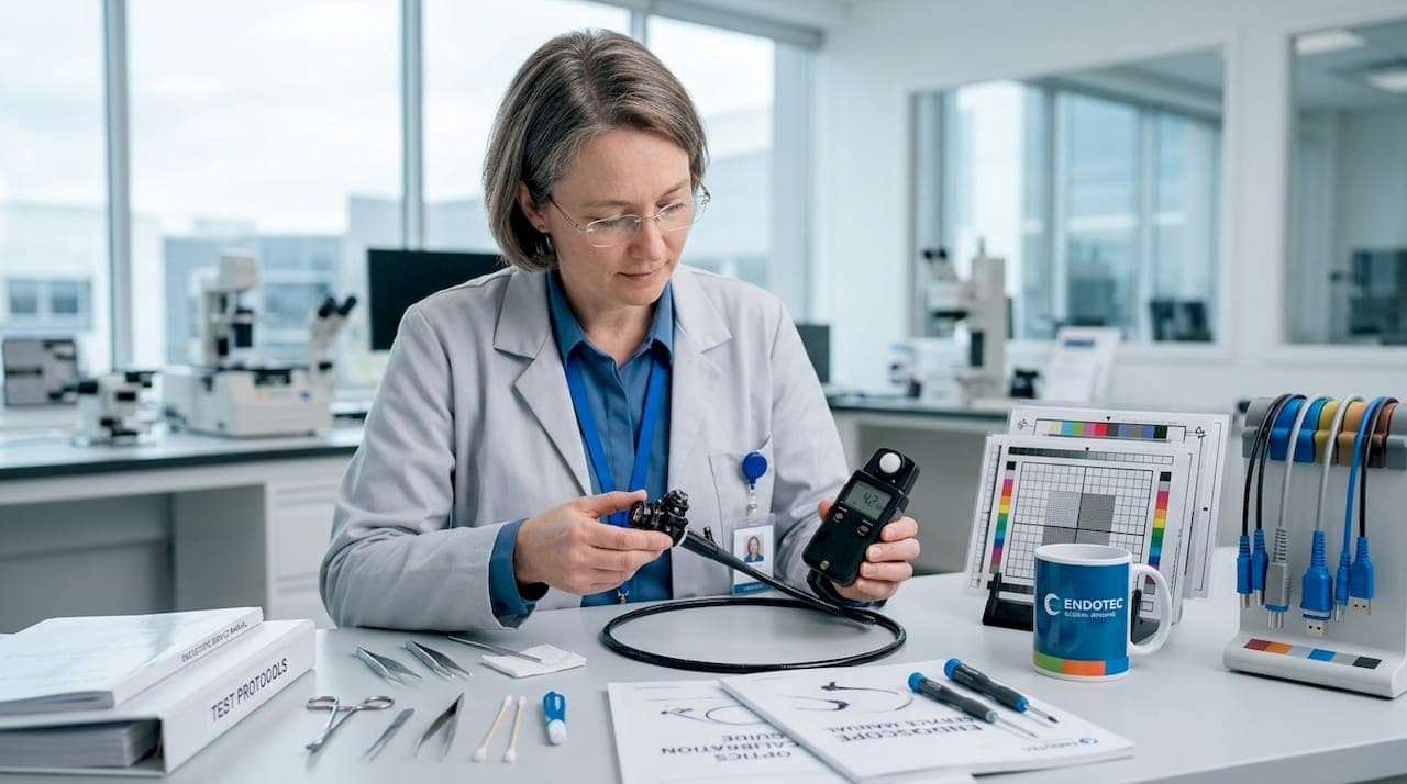
Explaining endoscope image quality is more complex than most professionals expect. The instinct is to look at pixel count and call it done. But veterinary clinicians trying to identify a mucosal lesion in an equine airway, or an NDT technician checking a turbine blade for micro-cracks, both know that a high-megapixel spec sheet means nothing if the image on screen is washed out, blurry, or distorted. True image quality is determined by an interconnected chain of factors: optics, illumination, digital processing, and how well your entire capture workflow holds together. This article breaks down each link in that chain with the specificity you actually need.
Table of Contents
- Key takeaways
- Optics and illumination: the foundation of image clarity
- Digital processing and encoding: where image fidelity lives or dies
- Endoscope types compared: how technology shapes what you see
- Practical steps for improving endoscope image quality
- My take on what actually determines endoscope image quality
- How 1800endoscope supports better imaging from scope to archive
- FAQ
Key takeaways
| Point | Details |
|---|---|
| Resolution alone is misleading | Pixel count matters less than lens quality, aberration control, and illumination consistency in real-world use. |
| Processing and encoding shape the final image | Digital capture pipelines and DICOM compliance directly affect sharpness, metadata preservation, and retrieval reliability. |
| Zoom degrades clarity without correction | High-magnification zoom causes contrast loss and ghosting unless adaptive aberration correction is applied. |
| Interoperability is a quality factor | Images that become orphaned due to poor standards implementation are effectively useless, regardless of their original clarity. |
| Maintenance protects optical performance | Routine cleaning and lens checks prevent contamination artifacts that degrade image quality over time. |
Optics and illumination: the foundation of image clarity
Most conversations about factors affecting endoscope images start and end with resolution. Resolution matters, but it is only one piece. What actually determines whether a veterinarian can distinguish healthy tissue from inflamed mucosa, or whether an inspector can resolve a 0.5mm crack in a casting, is a combination of lens quality, contrast, and light.
Resolution vs. spatial detail. Nominal pixel resolution describes how many points the sensor can capture. Spatial resolution describes the finest detail you can actually resolve in the image. These two numbers are not the same. Research on tethered capsule endomicroscopy demonstrates that spatial resolution at the micrometer scale provides far more clinically useful information than nominal pixel counts alone. A sensor with fewer pixels and a high-quality optical pathway will consistently outperform a high-megapixel system with poor lenses.
Lens quality introduces several variables that degrade image clarity before a single pixel is recorded:
- Chromatic aberration splits white light into color fringing at high-contrast edges, muddying fine detail
- Spherical aberration causes peripheral softness, which is especially problematic in wide-angle industrial borescope views
- Low contrast transmission flattens tissue or surface texture, reducing diagnostic value
- Light uniformity is critical: uneven illumination creates hotspots in the center and shadows at the edges, which is a common failure mode in poorly specified light sources
Zoom and focus compounds these issues. Liquid lens-based continuous zoom systems achieve impressive magnification ranges but face contrast degradation due to reduced light throughput and nonlinear lens aberrations. In a veterinary scope examining a trachea, this translates to a sharp center and soft, artifact-ridden periphery at maximum zoom.
Pro Tip: Before accepting any endoscope system, check the lens transmittance specification. Quality optics should transmit at least 90% of available light. Also verify that your light source output matches the scope's recommended illumination range. Mismatched illumination is one of the most common causes of poor image contrast in the field.
Digital processing and encoding: where image fidelity lives or dies
You can have flawless optics and still ruin image quality in the capture pipeline. This is the part of explaining endoscope image quality that most equipment buyers overlook until they have a workflow problem.
Here is how the digital pathway typically introduces quality loss:
- Analog-to-digital conversion at the sensor level introduces noise if the camera module is under-powered or thermally stressed during long procedures
- Video compression codecs like MPEG-2 or low-bitrate H.264 discard image data to reduce file size, causing visible compression artifacts in high-motion sequences
- Non-compliant capture software saves images in formats that strip metadata, making it impossible to associate an image with a patient record or inspection report
- Export and format conversion between proprietary systems and clinical or inspection software introduces additional generation loss
The last two points connect directly to DICOM compliance. DICOM is the international standard for medical image encoding and transmission, and it governs not just file format but the rich metadata that ties each image to a procedure, date, operator, and patient. Systems that treat DICOM as a secondary feature, rather than a native capability, create orphaned images and metadata mismatches that destroy traceability and make quality review nearly impossible.
This matters for industrial inspection just as much as for veterinary practice. When an NDT technician documents a weld inspection, images that cannot be reliably retrieved and associated with a specific component serial number are worthless for compliance or re-inspection.
AI-assisted video reporting systems underscore the stakes. Automated reporting accuracy of 79 to 83% in clinical endoscopy settings is directly tied to consistent frame clarity and image quality. Degraded or inconsistently captured footage causes these systems to fail or produce unreliable outputs.
Pro Tip: When selecting a recording or capture system, ask specifically whether DICOM is native to the software architecture or added as a plugin. Native DICOM compliance preserves full metadata fidelity from the moment of capture, not just at export. This is the difference between images you can always retrieve and images that periodically disappear from your records.
Endoscope types compared: how technology shapes what you see
Understanding endoscope visual fidelity also requires understanding how much the device type itself shapes image quality. Not all endoscopes are built to the same optical specifications, and the gap between device categories is wider than many buyers realize.
| Endoscope type | Typical resolution | Illumination | Zoom capability | Best application |
|---|---|---|---|---|
| Rigid veterinary scope | Very high (direct optics) | LED or fiber | Fixed or limited | Small animal surgery, arthroscopy |
| Flexible fiber-optic | Moderate (fiber bundle) | Fiber light guide | Minimal | Equine airway, GI tract access |
| Flexible videoscope | High (CCD/CMOS sensor) | LED tip | Digital zoom | Veterinary diagnostics, detailed GI work |
| Industrial borescope (rigid) | High (direct optics) | LED array | Fixed | Pipe, engine, and weld inspection |
| Industrial videoscope (flexible) | High (CMOS sensor) | LED tip | Digital/optical | Turbine, aerospace, NDT inspection |
Rigid endoscopes carry light and image data through a direct optical pathway, which preserves the highest native resolution and contrast. The tradeoff is access: rigid designs cannot navigate bends. Flexible fiber-optic scopes trade optical quality for reach, with the fiber bundle itself introducing a characteristic honeycomb pattern artifact that limits resolving power.
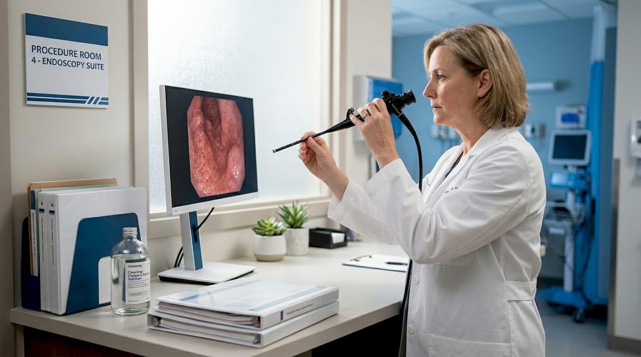
Flexible videoscopes place the sensor at the tip of the scope, bypassing the fiber bundle entirely. This significantly improves image clarity for veterinary GI work and detailed airway inspection in large animals. Working distance also plays a major role. Scopes designed for close-contact tissue examination operate at 5 to 20mm, where their optics are optimized. Pulling back beyond that range softens the image in ways that are not compensated by digital zoom.
Advanced aberration correction technology is narrowing the quality gap at high zoom. Physics-guided neural network methods can now correct dynamic lens aberrations in real time during continuous zoom, maintaining contrast and sharpness at magnifications that would previously produce unusable images. This technology is beginning to appear in premium videoscope systems and will likely become standard in the next generation of diagnostic scopes.
Practical steps for improving endoscope image quality
Knowing what influences endoscope imaging is only useful if it translates to what you do before, during, and after a procedure. Here are the best practices for endoscope quality that consistently make the biggest difference in the field.
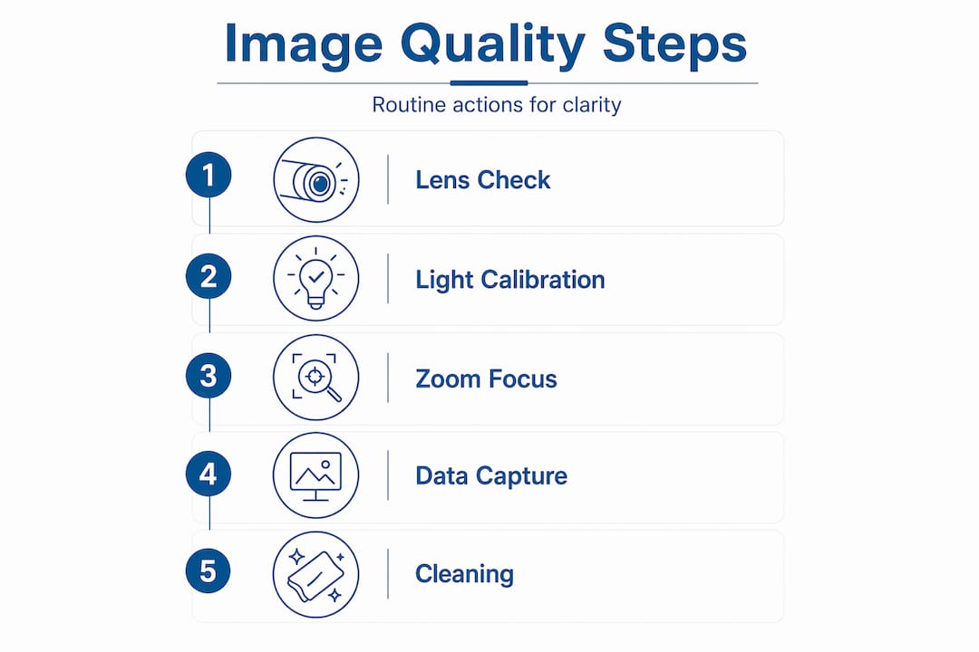
Routine optical checks. Before every session, visually inspect the objective lens for debris, smearing, or moisture. A contaminated distal tip degrades image quality far more than any camera limitation. For fiber-optic scopes, check for broken fiber bundles, which appear as dark spots that reduce effective resolution.
Illumination calibration. Match your light source intensity to the working distance of the procedure. Veterinary airway scopes working at 20 to 50mm need different illumination levels than industrial scopes inspecting wide-bore pipes. Most modern LED light sources allow adjustable output, and calibrating this setting at the start of a session prevents both washout and underexposed detail.
Environment and contrast adjustment. In industrial settings, surface reflectivity matters. Highly polished metal surfaces cause specular reflections that blow out image detail. Reduce light intensity or increase working angle to recover surface texture. In veterinary work, biological fluids can coat the lens mid-procedure. A brief water flush through the scope's irrigation channel restores clarity without withdrawing the instrument.
- Verify that your video capture format is set to the highest available resolution before starting recording
- Avoid converting video files through multiple format changes before archiving
- Store images in DICOM or another lossless format whenever your workflow supports it
- Integrate with PACS systems for reliable image retrieval and audit trails
Cleaning and maintenance. Enzymatic cleaning of flexible scopes removes protein deposits that gradually cloud the optical pathway. Skipping or shortening this step is one of the most common sources of progressive image degradation in busy veterinary and industrial operations.
Pro Tip: Confirm device compatibility across your entire workflow before purchasing. A videoscope that outputs via a proprietary connector and requires a format conversion to reach your practice management software introduces at least one generation of quality loss at every step. Systems designed with open standards throughout produce consistently better final images.
My take on what actually determines endoscope image quality
I've worked with enough veterinary clinics and industrial inspection teams to say this clearly: the obsession with megapixel counts is the single most misleading factor in endoscope purchasing decisions. I've seen facilities operate a lower-resolution scope with excellent glass and a well-matched light source and get diagnostic images that outperform "superior" specs systems running through a poor capture pipeline.
The hidden problem I encounter repeatedly is interoperability failure. A clinic invests in a high-quality videoscope, pairs it with capture software that was never designed to be natively DICOM-compliant, and then discovers months later that hundreds of procedure images have no patient association. The images looked fine on screen. They were useless in the record. That is an image quality failure just as real as a blurry lens.
What I've found actually works is thinking about image quality as a system, not a spec. Optics, illumination, processing, encoding, and data exchange all contribute to what you can see and what you can prove you saw. When one element in that chain is weak, the others cannot compensate fully. The practices that get this right invest in scopes with quality glass, match their light sources properly, use natively compliant capture software, and run regular maintenance programs. They rarely have image quality complaints. The ones chasing spec sheets and ignoring workflow integration are the ones calling us with problems.
— Endoscope
How 1800endoscope supports better imaging from scope to archive
At 1800endoscope, we build our product recommendations around the same holistic framework described throughout this article, because we know that optics, illumination, and workflow all have to work together.

For veterinary professionals, the portable 6mm airway videoscope delivers direct-monitor SD card video recording in a system designed for field-ready reliability. For equine practitioners needing clear upper airway imaging, the 8mm USB airway field scope pairs a quality distal sensor with a rugged build for barn-side use. Industrial inspection teams can browse the full range of NDT borescopes and flexible videoscopes at 1800endoscope.com, where every system is selected for real-world optical and capture performance. Whether your priority is veterinary diagnostics or precision industrial inspection, our catalog covers the full spectrum of imaging needs at pricing that works for clinics and field teams alike.
FAQ
What is explaining endoscope image quality, really?
Explaining endoscope image quality means accounting for every factor in the imaging chain: lens optics, illumination, digital processing, encoding standards, and workflow interoperability. Pixel count alone is not an adequate explanation.
Why does DICOM compliance affect image quality?
DICOM standards preserve image metadata and prevent orphaned files, which means images remain accessible, attributable, and usable for diagnostics or inspection records long after capture.
How does zoom affect endoscope image clarity?
Increasing zoom reduces light throughput and introduces dynamic lens aberrations, causing contrast loss and ghosting at high magnifications. Advanced aberration correction systems can compensate, but standard scopes show measurable quality degradation at maximum zoom.
What is the best practice for maintaining image quality daily?
Clean the objective lens and distal tip before every procedure, calibrate light source intensity to your working distance, and verify that your capture format is set to maximum resolution. These three steps prevent the most common sources of image degradation in both veterinary and industrial settings.
Do flexible and rigid endoscopes differ significantly in image quality?
Yes. Rigid scopes use direct optical pathways that preserve higher native contrast and resolution. Flexible fiber-optic scopes introduce honeycomb artifacts from the fiber bundle, while flexible videoscopes place the sensor at the tip to recover much of that lost clarity.
Recommended
- Endoscope Endoscopy FAQ and Inspection Procedures, Endoscopy Museum!
- History of Endoscopy Endoscopes Endoscope Museum Olympus� Pentax� Fujinon� ACMI�
- Pre-Owned Olympus� Pentax� Fujinon� Endoscopes, Endoscopy Systems 1800Endoscope.com
- Olympus� Pentax� Fujinon� Storz Welch Allyn Video Endoscopy Tower Systems! 1800Endoscope.com