
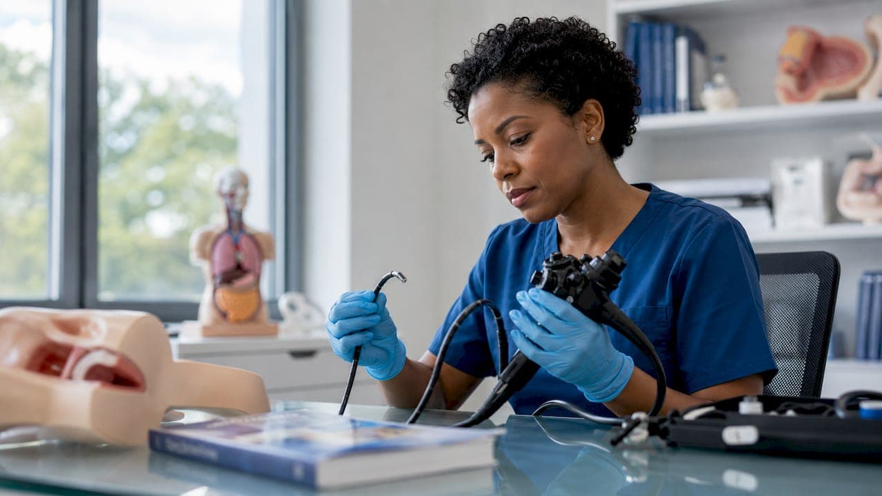
An endoscope is defined as an integrated optical and mechanical inspection instrument that combines a lens system, illumination source, control mechanism, and working channels into a single device for internal visualization. Explaining endoscope working parts requires more than naming components. Each part performs a specific mechanical or optical function, and failure in any one of them compromises the entire diagnostic outcome. The two primary design families are flexible and rigid endoscopes, each built from distinct materials and suited to different anatomical or industrial pathways. Endoscopes employ illumination sources, optical fibers, and camera sensors to deliver the high-resolution images that make minimally invasive diagnosis and intervention possible.
What are the major physical components of an endoscope?
The insertion tube is the most mechanically complex section of any endoscope. Its construction uses layered polymer sheaths with embedded mesh structures that balance chemical resistance, torque transmission, and flexibility for the anatomy it targets. The outer sheath resists body fluids and disinfectants, while the inner braid transmits rotational force from the operator's hand to the distal tip.
The distal tip is the business end of the instrument. It houses the objective lens or CMOS/CCD image sensor, illumination nozzles, and the openings for working channels. The micro-machined distal tip housing combines optical, mechanical, and fluid handling components engineered for durability through repeated disinfection and clinical use. Every millimeter of space in this housing is occupied by a functional element.
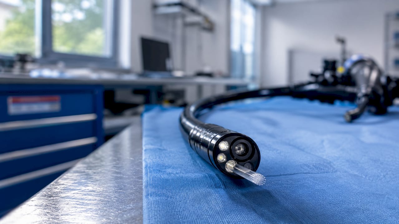
The control handle sits at the proximal end and gives the operator directional authority over the distal tip. Flexible endoscopes steer the distal tip up to 180 degrees in multiple directions using high-tensile angulation wires linked to manual control dials. That range of motion is what allows a flexible scope to navigate curved anatomy without tissue trauma.
Working channels run the full length of the insertion tube and serve three distinct functions:
- Biopsy channel: Passes forceps, cytology brushes, or retrieval baskets to the target site
- Suction channel: Clears fluid and debris from the field of view during the procedure
- Irrigation channel: Delivers water or saline to wash the lens or distend a cavity
All working channels use PTFE lining to reduce friction and resist chemical degradation. The light guide bundle runs alongside these channels, transmitting illumination from an external or integrated light source to the distal tip.
Pro Tip: When selecting a flexible endoscope for a new application, check the working channel diameter first. A channel too narrow for your intended instruments forces a scope replacement, not just an accessory swap.
How do optical and illumination systems produce clear internal images?
The optical path in an endoscope determines image sharpness, color accuracy, and field of view. Rigid endoscopes use relay lens systems of glass rod lenses for image transmission, while flexible endoscopes rely on coherent fiber-optic bundles or tip-mounted digital sensors like CMOS or CCD chips. The distinction matters because each method carries different trade-offs in resolution, cost, and repairability.
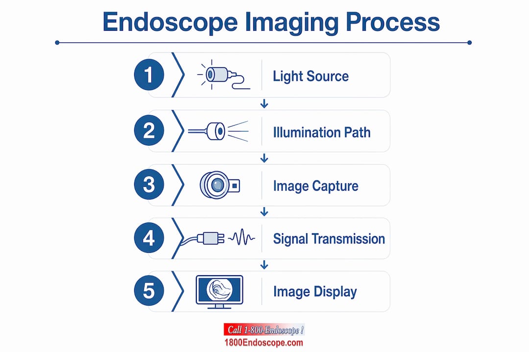
Fiber-optic bundles transmit images through thousands of aligned glass fibers. Each fiber carries one pixel of information, so the total fiber count sets the image resolution ceiling. Higher numerical aperture in fiber optics increases light collection but introduces subtle image fuzziness at the edges. Tip-mounted CMOS sensors bypass this trade-off entirely by digitizing the image at the source, which is why video endoscopes consistently outperform fiber-optic scopes in resolution.
| Optical method | Image quality | Repairability | Typical application |
|---|---|---|---|
| Glass rod relay lens | Highest clarity | Moderate | Rigid surgical scopes |
| Coherent fiber bundle | Good, pixel-limited | Low | Flexible diagnostic scopes |
| Tip-mounted CMOS/CCD | Excellent, HD capable | Low to moderate | Video endoscopes |
Light source technology directly affects tissue color rendering. LED sources offer long service life and consistent color temperature. Xenon sources produce a broader spectrum closer to daylight, which improves tissue differentiation. Laser-based illumination is used in narrow-band imaging applications where specific wavelengths enhance vascular contrast. Choosing the right light source for your scope type and procedure changes what you can see, not just how bright the image appears.
Bending the insertion tube stresses the fiber bundle. Repeated sharp bends break individual fibers, creating black spots in the image. These spots are irreversible. Keeping bend radius within the manufacturer's specification during storage and use is the single most effective way to extend fiber bundle life.
Pro Tip: Before each procedure, hold the scope against a light source and inspect the fiber bundle output for dark spots. A scope with more than a few broken fibers will degrade image quality below diagnostic threshold.
What vulnerabilities affect endoscope parts and their lifespan?
Component failure in endoscopes follows predictable patterns. Knowing where failures originate lets you build a maintenance program that catches problems before a scope fails mid-procedure.
- Distal tip seal failure: Fluid ingress into CMOS/CCD sensors is the leading cause of total endoscope failure. A compromised seal allows disinfectant or body fluid to reach the imaging electronics, which causes irreversible damage.
- Angulation wire fatigue: Wire fatigue causes loss of tip control during procedures. The bending section uses interlocking rings controlled by Bowden cables, and repeated articulation degrades wire tension over time.
- Insertion tube sheathing degradation: High-level disinfection agents attack the outer polymer layer. Micro-cracks in the sheath allow fluid ingress deeper into the tube structure.
- Working channel occlusion: PTFE-lined channels are susceptible to scoring and bioburden buildup despite thorough cleaning. Scored channel walls trap protein residue that standard brushing cannot remove.
- Light guide bundle breakage: Fiber breaks accumulate silently until image quality drops below acceptable levels, often without a clear triggering event.
Maintaining the airtight seal on the distal tip prevents the most costly endoscope failures. Current precision manufacturing improvements focus specifically on this seal because no amount of post-failure repair restores a water-damaged imaging sensor to full function. Leak testing after every procedure is the standard that separates facilities with low repair costs from those with chronic scope failures.
Professional endoscopy guidelines emphasize consistent maintenance and timely parts replacement to preserve device integrity. Leak testers, channel brushes, and visual inspection protocols are not optional steps. They are the difference between a scope that lasts years and one that fails within months. For practical field guidance, the endoscope inspection best practices resource covers current 2026 protocols in detail.
How do rigid and flexible endoscope designs differ in structure and use?
Rigid and flexible endoscopes solve the same visualization problem through fundamentally different engineering approaches. The choice between them is determined by anatomy, not preference.
Rigid endoscopes use stainless steel or titanium shafts that provide a perfectly straight optical path for relay lens systems. That straight path is non-negotiable for rod lens optics because any deviation scatters light and degrades the image. Rigid scopes deliver superior image clarity and are easier to sterilize because they have no bending section or angulation wires to trap contamination.
Flexible endoscopes use multi-layered polymer conduits and transmit images through fiber bundles or tip-mounted sensors. Balancing flexibility with durability is the central engineering challenge of flexible scope design. A scope that bends freely through a colon or a horse's airway must also resist the mechanical stress of thousands of articulation cycles without losing tip control.
| Feature | Rigid endoscope | Flexible endoscope |
|---|---|---|
| Shaft material | Stainless steel or titanium | Layered polymer with mesh braid |
| Image transmission | Glass rod relay lenses | Fiber bundle or CMOS/CCD sensor |
| Tip maneuverability | Fixed, no articulation | Up to 180 degrees in multiple planes |
| Sterilization | Autoclave compatible | High-level disinfection only |
| Best application | Straight-path surgery, arthroscopy, cystoscopy | GI endoscopy, airway inspection, industrial NDT |
Industrial NDT borescopes follow the same design logic. A rigid borescope inspects a straight engine cylinder bore with optical precision. A flexible borescope navigates a turbine blade cavity or a curved pipe run where a rigid shaft cannot reach. Understanding flexible borescope diagnostics helps technicians select the right tool for each inspection geometry.
Key Takeaways
Endoscope performance depends entirely on the integrity of each working part, from the distal tip seal to the angulation wires and optical system.
| Point | Details |
|---|---|
| Distal tip seal is critical | Fluid ingress through a failed seal is the leading cause of total endoscope failure. |
| Optical method determines resolution | Tip-mounted CMOS sensors outperform fiber bundles in image quality for video endoscopy. |
| Angulation wire fatigue is predictable | Repeated articulation degrades wire tension; monitor tip responsiveness at every use. |
| Rigid vs. flexible is anatomy-driven | Rigid scopes suit straight-path procedures; flexible scopes navigate curved anatomy. |
| Maintenance prevents most failures | Leak testing and channel inspection after every procedure extend scope lifespan significantly. |
What I've learned from watching scopes fail in the field
The professionals who get the most life from their endoscopes are not the ones with the most expensive equipment. They are the ones who understand what each component actually does and where it fails first.
The distal tip seal gets the least attention during routine use and causes the most expensive repairs. I've seen facilities skip leak testing for weeks at a time, then face a complete sensor replacement that costs more than a new entry-level scope. The math is not complicated. A leak tester costs a fraction of a sensor repair, and the test takes under two minutes.
Angulation wire fatigue is the failure mode that surprises operators the most. The scope steers normally right up until it doesn't. The degradation is gradual, which means it often goes unnoticed until tip control becomes unreliable during a procedure. Tracking tip responsiveness at each use, not just during scheduled maintenance, catches this early.
The rigid versus flexible decision deserves more deliberate thought than it typically gets. Rigid scopes are not inferior to flexible scopes. They are superior for the procedures they are designed for, and their optical clarity and sterilization compatibility make them the right choice in many surgical contexts. Defaulting to flexible scopes for every application is a procurement mistake that costs image quality and increases maintenance complexity.
The future of endoscope engineering points toward single-use distal tips with reusable handles, which eliminates the seal failure problem entirely for high-risk procedures. Professionals who understand component vulnerabilities now will be better positioned to evaluate those hybrid designs as they reach the market.
— Endoscope
Advanced endoscope systems available at 1800endoscope
1800endoscope stocks a full range of flexible and rigid endoscope systems built for veterinary, medical, and industrial diagnostic applications.

The portable airway inspection system offers HD video recording and SD card storage in a field-ready package suited for equine and small animal airway work. For facilities that need a broader selection, the complete endoscope catalog covers everything from small-diameter videoscopes to industrial NDT borescopes, along with replacement components, light sources, and leak testing accessories. 1800endoscope's team can help you match the right scope design and accessories to your specific diagnostic requirements.
FAQ
What is the most failure-prone part of a flexible endoscope?
The distal tip seal is the most failure-prone component. Fluid ingress through a compromised seal damages the CMOS or CCD imaging sensor and is the leading cause of total scope failure.
How does tip angulation work in a flexible endoscope?
Control dials in the handle pull high-tensile angulation wires that run through the bending section, steering the distal tip up to 180 degrees in multiple planes for navigation through curved anatomy.
What is the difference between fiber-optic and video endoscopes?
Fiber-optic scopes transmit images through coherent glass fiber bundles, with resolution limited by fiber count. Video endoscopes use a tip-mounted CMOS or CCD sensor to digitize the image at the source, producing higher resolution output.
Why are working channels a maintenance concern?
PTFE-lined working channels can develop scoring and harbor bioburden despite high-level disinfection. Scored channel walls trap protein residue that standard brushing cannot fully remove, creating infection control risks.
When should you choose a rigid endoscope over a flexible one?
Choose a rigid endoscope for straight-path procedures like arthroscopy, cystoscopy, or laparoscopy, where its glass rod relay lens system delivers superior image clarity and the shaft is compatible with autoclave sterilization.