
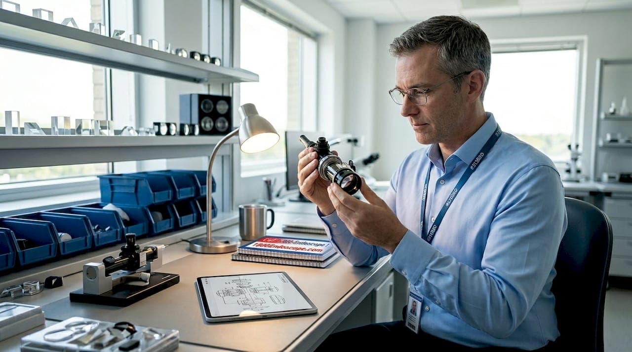
The role of optics in endoscopes is to guide light and form high-resolution images that enable precise internal visualization in medical, veterinary, and industrial applications. Every component in the optical chain, from objective lenses and relay prisms to light guides and distal-tip microlenses, directly determines what you see and how accurately you can act on it. Optical design decisions control field of view, image brightness, color fidelity, and resolution before a single pixel reaches your display. Understanding how these components interact is the difference between a diagnostic-grade image and one that leaves you guessing. This guide covers the optical components in endoscopes that matter most, with specific attention to veterinary and industrial contexts where image quality directly affects outcomes.
How do lenses and prisms influence image quality in endoscopes?
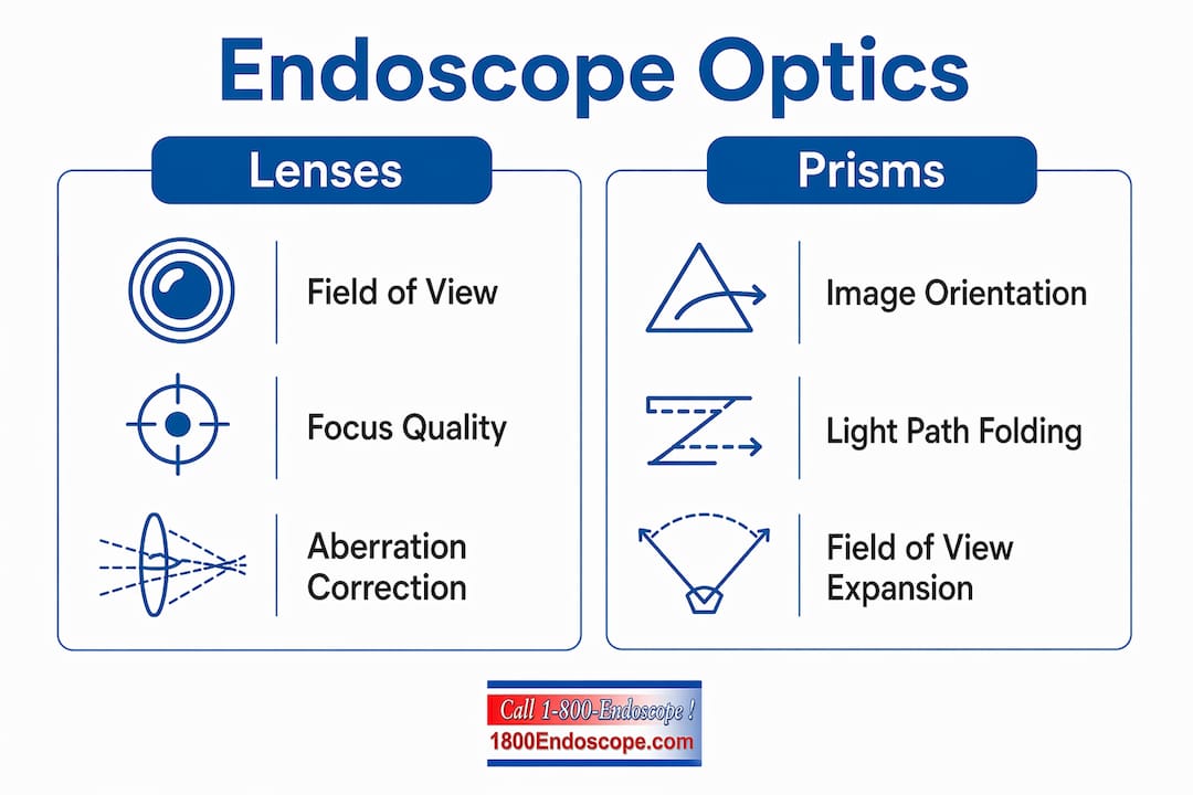
Objective lenses define the field of view and focus the image onto the sensor or fiber bundle at the distal tip. The geometry of these lenses sets the working distance, depth of field, and initial image magnification. A poorly specified objective lens cannot be rescued by downstream processing, which is why optical design starts here.
Prisms are core components controlling image orientation, light-path folding, and field of view in endoscopes. They correct the natural inversion that occurs when light passes through a lens system, and they fold the optical path to keep the scope diameter small enough for clinical or industrial use. Without prisms, a rigid endoscope would need to be physically longer to achieve the same optical path length, making it impractical for most procedures.
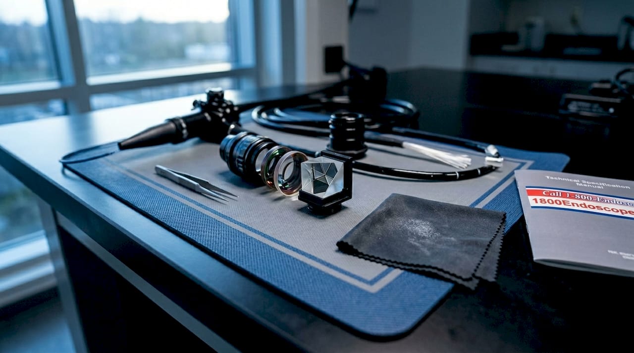
Prism geometry directly affects how much of the scene you capture. Wide-angle prisms expand the field of view but introduce aberrations that must be compensated elsewhere in the system. Narrow-angle prisms trade field of view for depth resolution, which suits applications like targeted biopsy or precision industrial inspection. Multi-prism systems use sequential elements to compensate chromatic aberration, where different wavelengths of light refract at slightly different angles and produce color fringing at image edges.
| Prism type | Field of view | Best application |
|---|---|---|
| Wide-angle prism | Broad, 120°+ | General survey, large-cavity veterinary scopes |
| Narrow-angle prism | Focused, 60° or less | Targeted biopsy, industrial defect localization |
| Multi-prism system | Variable, aberration-corrected | High-color-fidelity diagnostic endoscopes |
Pro Tip: When specifying a scope for equine or large-animal work, confirm the prism configuration supports the field of view you need at the working distance typical for that anatomy. A wide-angle prism that performs well in a bovine rumen will produce edge aberrations that compromise detail in a narrow equine airway.
What role does distal-tip aberration correction play in imaging performance?
Miniaturized fiber endoscopes face a specific optical challenge: the smaller the distal tip, the harder it is to correct for the aberrations that degrade resolution and color accuracy. Standard lens fabrication methods cannot produce the freeform surface geometries needed at sub-millimeter scales, which is why distal-tip optics have historically been the weakest link in fiber-based systems.
Recent advances change that picture significantly. A microlens integrated on an imaging-fiber tip can achieve a 60-degree field of view and 7.13 line pairs per millimeter resolution, enabled by freeform surfaces fabricated via two-photon polymerization. That resolution figure is meaningful: it represents the threshold at which fine mucosal texture and early-stage lesions become distinguishable rather than blurred.
The practical implications for veterinary and industrial users are direct:
- Full-color imaging at the distal tip means you are not interpreting a grayscale or color-shifted image when assessing tissue health or surface defects.
- Higher resolution at the tip reduces the need to reposition the scope repeatedly to confirm findings, cutting procedure time.
- Aberration correction near the distal tip is the single most impactful upgrade path for fiber-based scopes used in small-animal or narrow-bore industrial inspections.
- Freeform microlens fabrication is not yet standard across all product lines, so asking suppliers specifically about distal-tip lens design is worth the conversation.
Pro Tip: For small-animal endoscopy where scope diameter is constrained to 2mm or less, prioritize systems that specify distal-tip aberration correction explicitly. A scope with a high-megapixel sensor but uncorrected distal optics will underperform a lower-resolution scope with a well-corrected tip lens.
How do illumination optics and numerical aperture affect image brightness?
Illumination is where many otherwise well-specified endoscope systems lose performance. The light guide carries energy from the external source to the distal tip, but the efficiency of that transfer depends entirely on how well the numerical aperture of the light guide matches the acceptance angle of the source and the delivery geometry at the tip.
Endoscopic light guides with optimized numerical aperture are essential for high light capture and color fidelity, significantly affecting image brightness and quality. NA determines the cone angle of light the fiber accepts. A mismatch between source NA and guide NA means a portion of the available light is simply rejected at the coupling interface, reducing brightness at the tip before the image is even formed.
Getting illumination right requires attention to four specific factors:
- NA matching at the source coupling. The light source output NA must not exceed the fiber bundle's acceptance NA, or you lose light at entry.
- Anti-reflection coatings on coupling optics. Each uncoated glass-air interface reflects approximately 4% of incident light. In a multi-element illumination path, uncoated surfaces compound into a measurable brightness penalty.
- Fiber bundle packing density and diameter uniformity. Inconsistent fiber diameters create hot spots and dark zones at the distal tip, producing uneven illumination that degrades contrast across the image.
- Polarization management. Certain tissue types and industrial surfaces produce specular reflections that wash out detail. Polarization filters in the illumination path reduce glare and improve surface contrast, particularly useful in wet veterinary environments or reflective metal inspection.
Suboptimal illumination components bottleneck image brightness even when distal imaging optics are high specification. This is the most common source of unexplained image quality complaints in the field. If your scope produces dim or color-shifted images despite a functioning light source, the illumination path optics are the first place to investigate. For guidance on selecting the right light source for your setup, the veterinary light source selection framework covers the key decision points in detail.
What advanced optical technologies support multi-modal and 3D endoscopic imaging?
Multi-modal imaging, specifically the ability to switch between white-light and near-infrared modes within a single procedure, requires objective lens designs that handle two distinct spectral ranges without producing misregistered images. The optical challenge is that glass refracts near-infrared light differently than visible light, so a lens optimized for white-light imaging will shift the focal point when switched to NIR mode.
A multi-region lens design can separate white light and near-infrared beams, minimizing positional displacement to produce sharp dual-mode images with high color reproducibility. This matters clinically because NIR imaging with fluorescent contrast agents reveals vascular patterns and lymphatic flow that white-light imaging cannot show. If the two image modes are spatially offset, overlay-based diagnostics become unreliable.
Stereoscopic 3D endoscopy introduces a separate set of optical constraints. Current dual-aperture systems provide depth perception through head-mounted displays, but current 3D endoscope setups lack quantitative 3D data recording, leading to subjective and variable visual estimation of lesion size. That limitation has real consequences for staging decisions in oncology and for dimensional inspection in industrial NDT work.
The emerging solution combines optical hardware with computational reconstruction:
- Spatio-angular disparity methods capture depth information from multiple viewpoints simultaneously, enabling post-procedure 3D mapping of surface morphology.
- For industrial borescope applications, this translates to quantitative defect sizing rather than operator-estimated measurements.
- NIR and white-light dual-mode scopes are increasingly relevant in equine and large-animal surgery, where perfusion assessment during soft-tissue procedures adds diagnostic value.
- Wavelength-dependent wavefront distortion limits broadband digital correction in lensless fiber systems, increasing complexity or reducing temporal resolution in multispectral imaging. Confirming spectral bandwidth requirements before specifying a digital lensless system avoids this constraint.
For industrial technicians, the NDT illumination accuracy considerations that apply to standard borescopes become even more critical when multi-modal imaging is involved, since illumination consistency directly affects the reliability of computational reconstruction.
How do optical design choices affect procedural efficiency and reliability?
Optical quality is not just a diagnostic metric. It directly affects how long a procedure takes and how much cognitive load it places on the operator. A stable, high-resolution image reduces the number of repositioning passes needed to confirm a finding, which shortens procedure time and reduces patient or equipment stress.
Robotic endoscope systems with stable imaging complete procedural tasks faster than manual instruments, and the optical quality of the imaging system is a primary driver of that advantage. The same principle applies to manual scopes: better optics reduce operator variability by making findings unambiguous rather than interpretive.
Key practical considerations for veterinary and industrial professionals include:
- Miniaturization trade-offs. Reducing scope diameter to access narrow anatomy or tight industrial spaces forces optical compromises. Understanding which compromises are acceptable for your specific application prevents purchasing a scope that cannot perform its primary task.
- Biopsy and defect targeting accuracy. A wide, well-corrected field of view allows the operator to position biopsy forceps or NDT probes accurately on the first pass. Distorted peripheral optics cause targeting errors that require multiple attempts.
- Operator fatigue and diagnostic consistency. Dim or color-inaccurate images require more active interpretation, increasing fatigue over long procedures and raising the risk of missed findings. Endoscope image quality factors that affect operator performance are well-documented and worth reviewing when evaluating new equipment.
Key takeaways
Optical system design, not camera resolution alone, determines endoscope imaging performance across veterinary, medical, and industrial applications.
| Point | Details |
|---|---|
| Prisms control image orientation | Prism geometry sets field of view and must be matched to the anatomy or inspection target. |
| Distal-tip aberration correction is critical | Freeform microlenses at the fiber tip enable full-color imaging and measurable resolution gains. |
| Illumination NA matching drives brightness | Mismatched numerical aperture at the source coupling reduces light delivery before the image forms. |
| Multi-modal lenses require displacement control | NIR and white-light modes must share a common focal plane to produce reliable overlay diagnostics. |
| Optical quality reduces operator variability | Stable, high-resolution imaging shortens procedures and lowers the risk of missed findings. |
Why the optical chain deserves more attention than the camera spec sheet
Most purchasing conversations in veterinary and industrial endoscopy start with sensor resolution and end there. That approach misses the point. I have seen high-megapixel videoscopes produce worse clinical images than older fiber systems because the distal optics were under-specified and the illumination path was assembled with mismatched components. The camera is the last element in a chain that starts at the light source coupling and runs through every glass surface to the sensor.
The most overlooked factor in field performance is light-guide quality. Technicians and clinicians routinely attribute poor image brightness to a failing light source when the actual problem is a degraded fiber bundle with broken fibers reducing transmission efficiency. Replacing the light source does not fix a compromised illumination path.
Distal-tip fabrication methods matter more than most product literature acknowledges. Two-photon polymerization enables freeform lens surfaces that conventional grinding cannot produce at sub-millimeter scales, and that difference is visible in the image. When evaluating fiber-based scopes for small-animal or narrow-bore industrial work, ask specifically how the distal microlens was fabricated and what aberration correction it provides.
The future of endoscope optics sits at the intersection of hardware precision and computational correction. Spatio-angular disparity methods and digital wavefront compensation will extend the performance of compact scopes beyond what pure hardware design can achieve. But those computational methods depend on a well-characterized optical foundation. Garbage in, garbage out applies to algorithms as much as it does to sensors.
— Endoscope
Explore endoscopes built around optical performance

At 1800endoscope, the catalog is built around professionals who need imaging performance they can rely on, not just a scope that fits the budget line. Whether you are specifying a small-diameter videoscope for feline gastrointestinal work, a flexible fiber scope for equine airway assessment, or an industrial NDT borescope for turbine blade inspection, optical component quality is the specification that determines whether your findings are actionable. The full endoscope catalog covers veterinary, medical, and industrial systems with advanced optical configurations, including HD videoscopes, fiber optic systems, and compatible light sources. For industrial inspection professionals, the borescope range includes options with multi-modal imaging capabilities suited to precision NDT applications.
FAQ
What are the main optical components in an endoscope?
The primary optical components in endoscopes are objective lenses, relay prisms, light guides, and distal-tip microlenses. Each element controls a specific aspect of image formation, from field of view and orientation to brightness and color fidelity.
How does numerical aperture affect endoscope image quality?
Numerical aperture determines the cone angle of light a fiber accepts, directly controlling how much light reaches the distal tip. A mismatch between source NA and light-guide NA reduces brightness and degrades color accuracy even when the imaging optics are high specification.
Why does distal-tip aberration correction matter for fiber endoscopes?
Uncorrected aberrations at the fiber tip blur fine detail and shift colors, making it difficult to distinguish tissue texture or surface defects. Freeform microlenses fabricated via two-photon polymerization correct these aberrations and enable full-color imaging at resolutions above 7 line pairs per millimeter.
What limits current stereoscopic 3D endoscopy systems?
Current dual-aperture 3D endoscope systems rely on head-mounted displays and cannot record quantitative 3D data, which means lesion size and defect dimensions are estimated visually rather than measured. Computational methods using spatio-angular disparity are emerging to address this limitation.
How do multi-modal endoscope optics handle NIR and white-light imaging?
Multi-region objective lens designs assign different refractive powers to separate spectral zones, minimizing the focal-point displacement between near-infrared and white-light modes. This keeps both image modalities spatially registered, which is required for reliable fluorescence overlay diagnostics.