
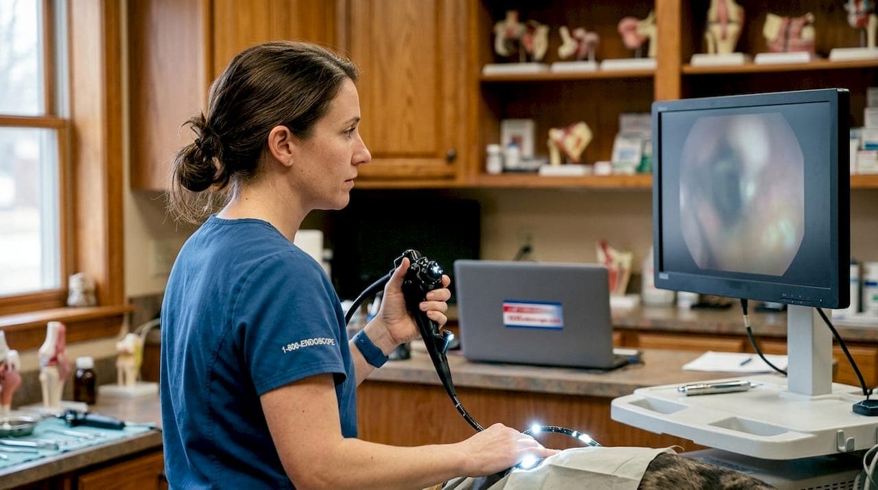
Shadow-free lighting in scopes is defined as a multi-source illumination system that eliminates cast shadows inside body cavities by overlapping light beams from multiple LED points arranged in a circular matrix. For veterinarians performing endoscopic examinations, this technology is the difference between a clear diagnostic image and a misleading one obscured by instrument shadows or anatomical obstructions. Understanding what is shadow-free lighting in scopes, how it works optically, and where it has real clinical limits gives you a sharper framework for choosing the right equipment. 1800endoscope builds this technology into its veterinary endoscope lineup specifically for in-cavity applications in small and large animal practice.
What is shadow-free lighting in scopes and how does the technology work?
Shadow-free lighting, also called shadowless illumination, works by placing multiple LED light sources in a circular array around the scope's distal tip. Each source emits a beam aimed at the same target zone. The beams overlap, so any shadow cast by one source is filled in by light from the adjacent sources.
The physics behind this is straightforward. A single light source creates a hard shadow with two zones: the umbra (the fully dark center) and the penumbra (the blurry edge). Multi-point LED arrays arranged in a circular matrix shrink the umbra and expand the penumbra until the shadow effectively disappears at the target surface. The result is a uniformly lit field with no dark pockets.
Collimating lenses are the component that makes this work cleanly. Each LED chip requires a precision collimating lens to convert its raw, scattered output into a directed beam. Without collimation, multiple LEDs produce overlapping halos and a distracting artifact called "ghosting," where several faint shadows appear instead of one clean image. Collimated beams converge mathematically at the tissue surface, producing a single, uniform column of light.
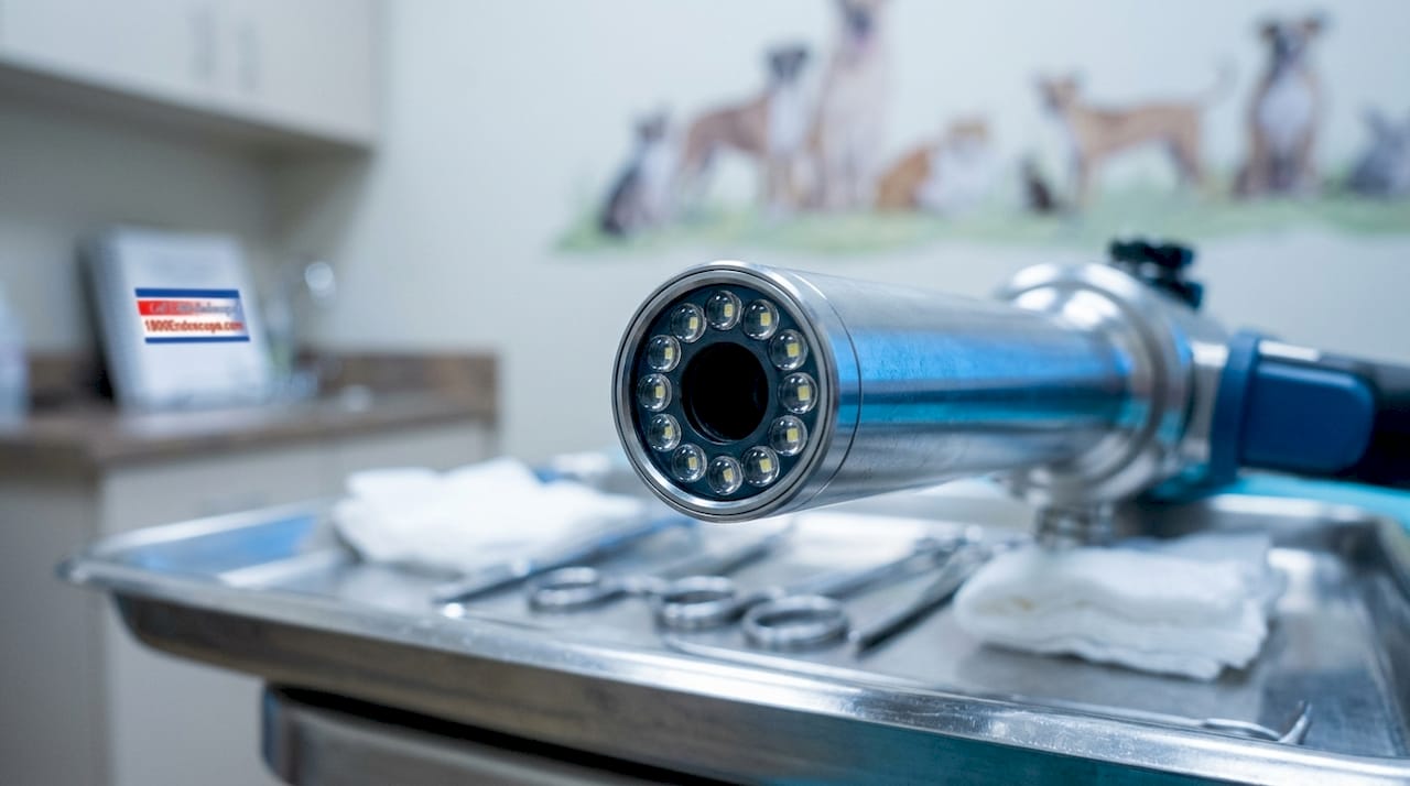
Pro Tip: When evaluating a veterinary scope's lighting specs, ask specifically whether each LED is paired with its own collimating lens. A scope with 12 LEDs but no collimation optics will produce ghosting artifacts that undermine the shadow-free benefit entirely.
Single-point light sources, including older fiber optic bundles fed from a remote lamp, cannot replicate this effect. They produce one beam with a fixed shadow geometry. Fiber optic bundles remain useful for transmitting high-intensity light from an external source, but the single-exit geometry means shadows from a biopsy forceps or the scope shaft itself will always fall on the tissue. For selecting light sources in endoscopic work, the number and arrangement of emitters matters as much as raw brightness.
What are the clinical benefits of shadow-free lighting for veterinary examinations?
The most direct benefit is improved diagnostic accuracy. When tissue surfaces are uniformly lit, color changes, lesions, and mucosal irregularities are visible without repositioning the scope to chase shadows. This is especially relevant in equine gastroscopy, where the large gastric volume and curved walls create complex shadow geometries with single-source lighting.
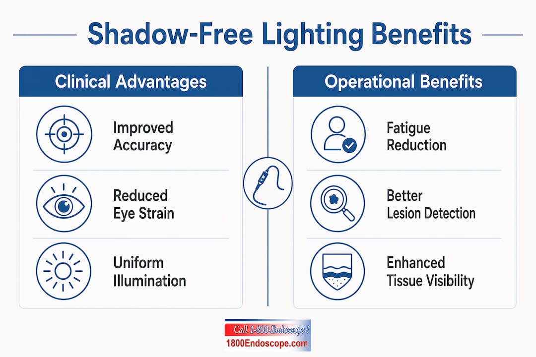
Consistent shadow-free illumination reduces eye strain and fatigue among clinicians by minimizing the high contrast between lit and shadowed areas during lengthy procedures. That reduction in contrast stress directly correlates with sustained precision. A veterinarian performing a 45-minute colonoscopy in a large dog benefits measurably from uniform light, because the visual system does not have to continuously adapt between bright and dark zones.
The surgical data from human medicine translates directly to veterinary practice. Integrated cordless LED lighting reduces tissue damage by 70% and improves tissue integrity maintenance, according to surgeon reports from breast surgery applications. That figure reflects what happens when clinicians can see clearly without repositioning instruments. In veterinary soft tissue surgery, the same principle applies: better visibility means fewer inadvertent cuts and cleaner dissection planes.
Key clinical advantages include:
- Uniform mucosal visualization across curved surfaces without repositioning
- Reduced procedure time because the clinician is not adjusting scope angle to compensate for shadows
- Lower cognitive load during complex procedures, freeing attention for diagnostic interpretation
- Improved image capture quality for video recording and telemedicine consultations
For veterinary professionals exploring LED-based scope illumination, the shift from single-source to multi-point arrays represents a genuine improvement in diagnostic capability, not just a specification upgrade.
How does in-cavity lighting outperform overhead lights in deep examinations?
Overhead surgical lights face a geometric problem that no amount of brightness can solve. The light travels from meters away, and any instrument, hand, or anatomical fold between the lamp and the target creates a shadow. Overhead lights require frequent manual repositioning due to shadows from personnel or instruments, disrupting workflow and increasing clinician cognitive load. That interruption cost is real and cumulative across a full surgical day.
The inverse square law explains why in-cavity lighting wins in deep examinations. Light intensity drops with the square of the distance from the source. A lamp 1 meter away delivers one-quarter the intensity of the same lamp at 0.5 meters. Fiber optic or LED-integrated scopes emit light within centimeters of tissue rather than meters away, which means the tissue receives dramatically more usable light even from a lower-wattage source.
The table below compares the two approaches across the criteria that matter most in veterinary endoscopy:
| Criterion | Overhead lighting | In-cavity scope lighting |
|---|---|---|
| Shadow geometry | Fixed; blocked by instruments and anatomy | Eliminated at the distal tip |
| Intensity at tissue | Reduced by distance (inverse square law) | High; source is centimeters from target |
| Repositioning required | Frequent | None during scope advancement |
| Deep cavity access | Ineffective beyond a few centimeters | Full illumination at any depth |
| Clinician fatigue | Higher due to contrast adaptation | Lower due to uniform field |
Pro Tip: For equine airway or rectal examinations, always confirm that your scope's light source is integrated at the distal tip rather than transmitted through a long fiber bundle. Bundle length attenuates light output, and a 3-meter bundle can lose a significant portion of its initial intensity before it reaches the tissue.
In-cavity lighting is not a replacement for overhead surgical lights in open procedures. The two systems serve different geometries. For any procedure where the scope tip enters a body cavity, whether a trachea, esophagus, colon, or uterus, the in-cavity source is the only practical way to achieve true shadow-free illumination. For veterinary airway procedures, this distinction is clinically significant.
What are the limitations of shadow-free lighting in scope imaging?
Shadow-free illumination is not universally ideal. The same uniform light that eliminates diagnostic shadows also removes depth cues from the image. Depth cues in endoscopic imaging come from shadows cast across surface features. When those shadows disappear, subtle mucosal texture, polyp borders, and early erosions can appear flatter than they actually are.
Directional illumination highlights subtle surface irregularities that uniform shadow-free lighting may flatten out. This is well-established in machine vision and applies equally to veterinary endoscopic imaging. A small raised lesion on the gastric mucosa may be more visible under angled light than under a perfectly uniform field.
The practical implications for veterinary professionals include:
- Flat mucosal lesions such as early adenomas or subtle erosions may be easier to detect with a slight directional component in the lighting
- Texture-dependent diagnoses like fungal plaques or roughened mucosa benefit from controlled shadow casting
- Scope systems with adjustable LED segments allow the clinician to shift between uniform and directional modes, which is the most flexible solution
Brightness alone is insufficient for visibility. The optical system, including collimating lenses and LED arrangement, determines whether the light serves the diagnostic task. A well-designed scope integrates both uniform illumination for navigation and the option for directional emphasis during close inspection.
Segmented LED ring lights allow professionals to activate specific LED segments to cast controlled shadows when surface texture matters. This capability is increasingly available in advanced veterinary videoscopes and represents the direction the technology is moving.
Key Takeaways
Shadow-free lighting in veterinary scopes works because multi-point LED arrays with collimating lenses overlap beams to eliminate shadows inside body cavities, delivering uniform illumination that overhead lights cannot achieve at depth.
| Point | Details |
|---|---|
| Core technology | Circular LED arrays with collimating lenses overlap beams to eliminate umbra and penumbra shadows. |
| In-cavity advantage | Placing the light source inside the cavity bypasses the inverse square law penalty of distant overhead lights. |
| Clinical benefit | Uniform illumination reduces clinician eye strain and improves diagnostic accuracy during long procedures. |
| Known limitation | Shadow-free lighting flattens depth cues; directional lighting is preferable for detecting subtle surface textures. |
| Equipment selection | Confirm each LED is paired with a collimating lens; multi-segment scopes offer both uniform and directional modes. |
What I've learned from watching shadow-free lighting change veterinary endoscopy
The shift from single-source fiber optic illumination to integrated multi-LED arrays is one of the most underappreciated improvements in veterinary endoscopy over the past decade. Clinicians who have used both systems consistently report that the difference is not subtle. It shows up in procedure confidence, image quality, and the ability to catch lesions that would have been missed under a shadowed field.
What I find most interesting is the coming generation of autonomous lighting systems that use depth cameras to dynamically adjust multiple light modules and prevent shadows without any manual input. That technology exists in human surgical suites today. Its migration into veterinary endoscopic equipment is a matter of time, not possibility.
The practical advice I give to any veterinary clinic evaluating new endoscopy equipment is this: do not evaluate lighting by lumen output alone. Ask how many LEDs are at the distal tip, whether each has a collimating lens, and whether the system allows segment control for directional emphasis. Those three questions will tell you more about real-world diagnostic performance than any brightness specification on a data sheet.
The clinics that invest in proper in-cavity illumination now will spend less time repositioning scopes, produce better documentation images, and catch pathology earlier. That is a clinical and economic argument that holds across every species and every procedure type.
— Endoscope
Advanced lighting options at 1800endoscope
Veterinary professionals who want to put shadow-free illumination to work in their practice will find a range of options at 1800endoscope, from compact portable systems to full videoscope setups with integrated LED arrays at the distal tip.

The portable airway endoscope from 1800endoscope is a 6mm system with direct monitor output and SD card recording, built for airway and upper GI work in small animals where in-cavity lighting is non-negotiable. For clinics that need a broader selection, the veterinary rigid endoscopy catalog covers a full range of rigid scopes with optimized lighting configurations for diagnostics across species. Every system is designed to deliver the uniform, shadow-minimized illumination that accurate internal examinations require.
FAQ
What is shadow-free lighting in a veterinary scope?
Shadow-free lighting is a multi-point LED illumination system at the scope's distal tip that overlaps beams from a circular array to eliminate shadows inside body cavities. It gives veterinarians a uniformly lit field without dark zones caused by instruments or anatomy.
How does shadow-free lighting differ from standard fiber optic illumination?
Standard fiber optic illumination exits from a single point and casts fixed shadows from any obstruction in the light path. Shadow-free systems use multiple LED sources with collimating lenses to fill those shadows by overlapping beams from different angles.
Does shadow-free lighting work in deep cavities like the equine colon?
Yes. Because the light source is at the scope tip inside the cavity, it bypasses the distance penalty of overhead lights and delivers high-intensity, uniform illumination at depth regardless of how far the scope has advanced.
Are there situations where shadow-free lighting is not the best choice?
Directional lighting is preferable when detecting subtle surface textures, early mucosal lesions, or raised irregularities, because uniform light flattens depth cues that help identify those features. Scopes with segmented LED control offer both modes.
What should veterinarians check when evaluating scope lighting quality?
Confirm that each LED at the distal tip is paired with a collimating lens, check the number of LED emitters in the circular array, and ask whether the system supports segmented control for switching between uniform and directional illumination. These factors determine real diagnostic performance.