
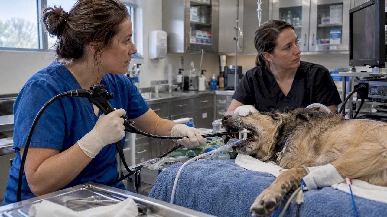
Endoscopy is the preferred first-line method for retrieving most esophageal and gastric foreign bodies in dogs and cats, with surgery reserved for cases where endoscopy fails or perforation is suspected. Any object lodged in the esophagus or airway needs same-day veterinary attention. Outcomes depend heavily on how quickly the animal is seen, what the object is made of, and whether the operator has experience with endoscopic retrieval.
TL;DR:
- Endoscopic removal has an 88% success rate in dogs and cats, with procedure times averaging about an hour, depending on the object and operator experience.
- Sharp objects, battery ingestion, or material ingested over a few hours should not be induced to vomit; prompt veterinary evaluation is crucial.
- Imaging with radiographs and ultrasound, plus contrast studies if needed, guide intervention by localizing the object and assessing for perforation.
- Appropriate tool selection, based on object shape and surface, is vital for efficiency and success during endoscopic retrieval.
- Surgery is reserved for failed endoscopy, objects beyond reach, or evidence of perforation, with hybrid techniques offering shorter anesthesia and hospitalization.
Table of Contents
- Recognizing the signs and what to do first
- Diagnostic workup: imaging before intervention
- Endoscopic retrieval: tools, technique, and when to stop trying
- Surgical removal and hybrid techniques
- Complications, prognosis, and what recovery looks like
- Preventing the next ingestion event
- What the evidence actually supports
- What equipment and training mean for retrieval outcomes
- Equipping your clinic for foreign body cases
- FAQ
- Sources
Recognizing the signs and what to do first
Dogs and cats rarely swallow something and show it clearly. Signs depend on where the object lodges and how long it has been there.
- Esophageal obstruction: repeated gagging, retching without producing vomit, drooling, reluctance to eat, or stretching the neck.
- Gastric or intestinal obstruction: vomiting (sometimes intermittent over days), lethargy, abdominal pain, reduced appetite, or a hunched posture.
- Respiratory involvement: coughing, gagging paired with labored breathing, or blue-tinged gums, which signal a possible airway object and need emergency care immediately.
- Red flags requiring urgent care: collapse, pale or tacky gums, a painful or distended abdomen, or known ingestion of a sharp object such as a fish hook or needle.
Do not induce vomiting if you suspect a sharp object, a battery, or something ingested more than a couple of hours ago. Withhold food and water until a veterinarian has examined the animal, since a full stomach complicates both sedation and endoscopic visualization. When you call the clinic, describe what was swallowed, roughly when it happened, and any material details (linear versus solid, metal versus fabric), because that history shapes whether the team reaches for endoscopy, imaging, or goes straight to surgery.
Pro Tip: Keep the packaging or a photo of the swallowed item if you have one. It helps the clinical team pick the right retrieval tool before the patient is even sedated.
Diagnostic workup: imaging before intervention
Plain radiographs are usually the first step, and most clinics will want at least two views (lateral and ventrodorsal) to localize the object and check for free abdominal gas, which suggests perforation. Radiographs show metal, bone, and dense plastic well but often miss radiolucent materials like fabric, rubber, or some types of plastic.
- Ultrasound adds value when radiographs are inconclusive, helping localize a mass, assess bowel wall thickening, and detect free fluid suggestive of perforation or peritonitis.
- Contrast studies using iodinated contrast are preferred when perforation is a concern, because leaked iodinated contrast is far less irritating to tissue than barium.
- Barium is avoided when perforation is suspected, and when it is used for diagnostic purposes, Merck Veterinary Manual guidance recommends waiting 24 hours before attempting endoscopy, since residual barium coats the mucosa and can damage endoscope channels.
- Diagnostic endoscopy itself is both a diagnostic and therapeutic step once imaging confirms an object is present and anesthesia is deemed safe.
One figure worth anchoring the whole workup on: endoscopic removal of esophageal and gastric foreign bodies succeeded in 88% of 92 cases in a retrospective review, which is why most clinics attempt endoscopy before considering surgery when the object's location and composition are endoscopy-compatible.
Logistics matter here too. Esophageal objects need fast scheduling since delay raises the risk of pressure necrosis and stricture, while a stable gastric foreign body sometimes allows a short window to arrange the right equipment and a fasted patient.

Endoscopic retrieval: tools, technique, and when to stop trying
Device selection is often the difference between a 20-minute retrieval and a failed attempt. Clinicians generally choose among a small set of tools based on the object's shape and surface:
- Alligator forceps grip irregular or textured surfaces well and are a common first choice for bone fragments or chewed objects.
- Rat-tooth forceps offer a firmer bite on dense material but can be less forgiving on friable or soft objects.
- Retrieval baskets suit smooth, rounded objects like balls or marbles that forceps tend to slip off.
- Snares work well on linear or elongated items, including some string or thread foreign bodies, though linear foreign bodies carry their own surgical considerations.
Positioning and airway protection matter as much as the tool itself, and understanding diagnostic steps like otoscopy and cytology for ear infections in dogs can inform related clinical assessments. For sharp objects such as fish hooks, passing a large endotracheal tube over the endoscope lets the clinician pull the object into the tube before withdrawal, protecting the esophageal mucosa from laceration on the way out. Insufflation should be used sparingly in a already-irritated esophagus, and most experienced endoscopists set a practical time limit on attempts, since a prolonged anesthetic episode without progress is itself a risk.
Operator experience is one of the strongest predictors of success. The same 92-case review found a mean extraction time of about an hour (https://pmc.ncbi.nlm.nih.gov/articles/PMC10538072/), with faster, more successful retrievals associated with more experienced endoscopists and with puppies specifically. Reviews of instrument use also note that certain device combinations, such as pairing a polypectomy snare with grasping forceps, have been associated with reduced extraction success in retrospective data, so matching one well-suited tool to the object tends to outperform stacking multiple devices.
Pro Tip: Document each retrieval attempt, including tool used, time elapsed, and visual findings. A clear record makes the decision to convert to surgery faster and more defensible if the first attempt doesn't work.
When an object cannot be safely grasped, keeps slipping, or the esophagus shows signs of deep ulceration or thinning, that's the signal to stop and move to surgical planning rather than repeating the same approach.

Surgical removal and hybrid techniques
Surgery becomes necessary when endoscopy fails, when the object has moved beyond endoscopic reach into the small intestine, or when imaging suggests perforation, in which case immediate surgical exploration takes priority over any retrieval attempt. ACVS guidance on gastrointestinal foreign bodies outlines gastrotomy (an incision into the stomach) and enterotomy (an incision into the intestine) as the standard surgical approaches, each carrying expected recovery and complication profiles that the surgical team will walk owners through before the procedure.
A hybrid approach, often called laparotomy-assisted or surgically-assisted endoscopy, has gained traction in selected cases. The surgeon opens the abdomen, then uses the endoscope to locate and manipulate the object from inside the stomach, sometimes avoiding a full gastrotomy incision altogether.
- Case-series data on this technique describe a five-case review where the assisted approach removed gastric foreign bodies while sparing a gastrotomy in selected patients.
- Comparative outcome data on linear foreign bodies found that laparotomy-assisted endoscopic removal was associated with shorter anesthetic time and hospitalization compared with standard gastrotomy, without a rise in major complications.
| Approach | Typical indication | Reported outcome pattern |
|---|---|---|
| Gastrotomy/enterotomy | Object inaccessible to endoscope or intestinal location | Standard recovery expectations per ACVS guidance |
| Laparotomy-assisted endoscopy | Abdomen already open, or endoscopy likely to fail | Reduced anesthetic time and hospitalization in comparative series |
| Standard endoscopy | Esophageal or gastric object reachable and graspable | 88% success in retrospective review |
The hybrid technique is not a replacement for gastrotomy in every case. It works best when the surgical team already has endoscopic equipment on hand and the object's size and position make endoscopic manipulation plausible.
Complications, prognosis, and what recovery looks like
Complication risk varies by procedure and by how long the object sat before removal.
- During endoscopic attempts: mucosal laceration, minor bleeding, or aspiration are the most common early complications, usually managed with supportive care and monitoring.
- Perforation is the complication that changes everything, since it requires immediate surgical repair rather than continued endoscopic effort.
- Later complications: esophageal stricture can develop over the following weeks if the mucosa was deeply damaged, which is why some patients need a recheck esophagoscopy during healing.
- Postoperative medical management commonly includes sucralfate to coat and protect irritated mucosa, acid-reducing medication such as a proton pump inhibitor, and appropriate pain control.
Most endoscopic retrieval patients go home the same day or after an overnight stay, with a short period of a bland or soft diet. Surgical patients typically need a longer hospital stay and activity restriction during incision healing. Any return of vomiting, regurgitation, or reluctance to eat after discharge warrants a prompt recheck rather than a wait-and-see approach.
Preventing the next ingestion event
Most foreign body cases are repeat behavior in predictable categories, which makes prevention genuinely actionable.
- Puppies and young dogs account for a large share of cases involving toys, socks, and bones, so rotate chew toys and supervise unstructured chewing time.
- Cats are especially prone to linear foreign bodies like string, ribbon, and dental floss, so these should be stored out of reach entirely rather than just out of sight.
- Households with small objects (coins, batteries, hardware) should store them in closed containers, since curious pica behavior in dogs doesn't discriminate by object type.
- After any ingestion event, keep the product packaging or a photo of the item and note the approximate time of ingestion, since that detail directly shapes the clinic's diagnostic plan.
What the evidence actually supports
The clinical picture is fairly consistent across recent literature: endoscopic removal succeeds in the large majority of appropriately selected cases, with one retrospective review reporting 88% success across 92 animals and mean extraction times near an hour. Hybrid surgical-endoscopic techniques show promise for shortening anesthesia and hospitalization in selected linear and gastric foreign body cases without raising complication rates.
What equipment and training mean for retrieval outcomes
Retrieval success depends as much on the operator's hands and judgment as on the scope itself, but the right tool shortens a difficult case considerably. Veterinary endoscopy equipment users report that a well-matched forceps or basket, paired with a trained eye, beats a more expensive scope used without practice. Clinics weighing new equipment are welcome to review our product documentation or reach out with procedural questions.
— Endoscope
Equipping your clinic for foreign body cases
A clinic doing endoscopic retrieval regularly needs a few things in place: a flexible scope with a working channel wide enough for the forceps or basket you intend to use, a diameter appropriate for the patient sizes you see, and enough portability that the system doesn't sit unused because it's awkward to set up. Smaller clinics and mobile practices often lean toward compact, monitor-type videoscope systems for exactly that reason.

Specialized suppliers stock the practical side of this workflow:
- Flexible scopes and accessories through our medical endoscope and accessory listings, including the New Portascope line.
- Retrieval-relevant forceps sized for small animal work on our biopsy forceps page.
- Rigid scope options for clinics that also handle arthroscopy or otoscopy cases, listed on our rigid endoscopy page.
- Repairs and general equipment sourcing through our main product and repair page.
For clinics building out a broader endoscopy capability, including borescope-style inspection tools used in some mixed-practice settings, our full catalog is the fastest way to compare options before reaching out to our team for specifics.
This article is general information, not a substitute for advice from a qualified doctor. Consult a qualified healthcare professional about your own circumstances before acting on anything here.
FAQ
What is the success rate of endoscopic foreign body removal in dogs and cats?
A retrospective review of 92 cases found endoscopic removal succeeded in 88% of animals, with success linked to patient factors and operator experience. Surgery remains the backup plan when endoscopic attempts fail or the object is beyond the scope's reach.
How long does endoscopic foreign body retrieval usually take?
The same case review reported a mean extraction time of about an hour(https://pmc.ncbi.nlm.nih.gov/articles/PMC10538072/), though individual cases ranged widely depending on the object and its location. Clinicians generally set a practical time limit and convert to surgery rather than extending a single attempt indefinitely.
What's the difference between alligator and rat-tooth forceps for retrieval?
Alligator forceps grip irregular or textured surfaces, making them a common choice for bone fragments or chewed material, while rat-tooth forceps hold dense objects firmly but can be less forgiving on soft or friable items. The choice depends on the object's shape and surface rather than one tool being universally better.
When does a foreign body case require surgery instead of endoscopy?
Surgery is needed when the object has moved beyond endoscopic reach, when imaging suggests perforation, or when endoscopic attempts fail to grasp or safely extract the object. In some gastric cases, laparotomy-assisted endoscopy can avoid a full gastrotomy while still using the abdomen's open access, per comparative outcome data.
Can I safely wait before taking my pet to the vet after they swallow something?
Esophageal or airway objects need same-day emergency evaluation, since delay raises the risk of tissue damage and stricture formation. Gastric foreign bodies sometimes allow a short window to reach a clinic, but any vomiting, gagging, or lethargy should be treated as urgent rather than watched at home.
Sources
- Endoscopic Retrieval of Esophageal and Gastric Foreign Bodies in Cats and Dogs: A Retrospective Study of 92 Cases
- Comparative outcomes following laparotomy-assisted endoscopic removal or gastrotomy for treatment of linear foreign bodies in dogs and cats.
- ACVS: Gastrointestinal foreign bodies (small animal)