
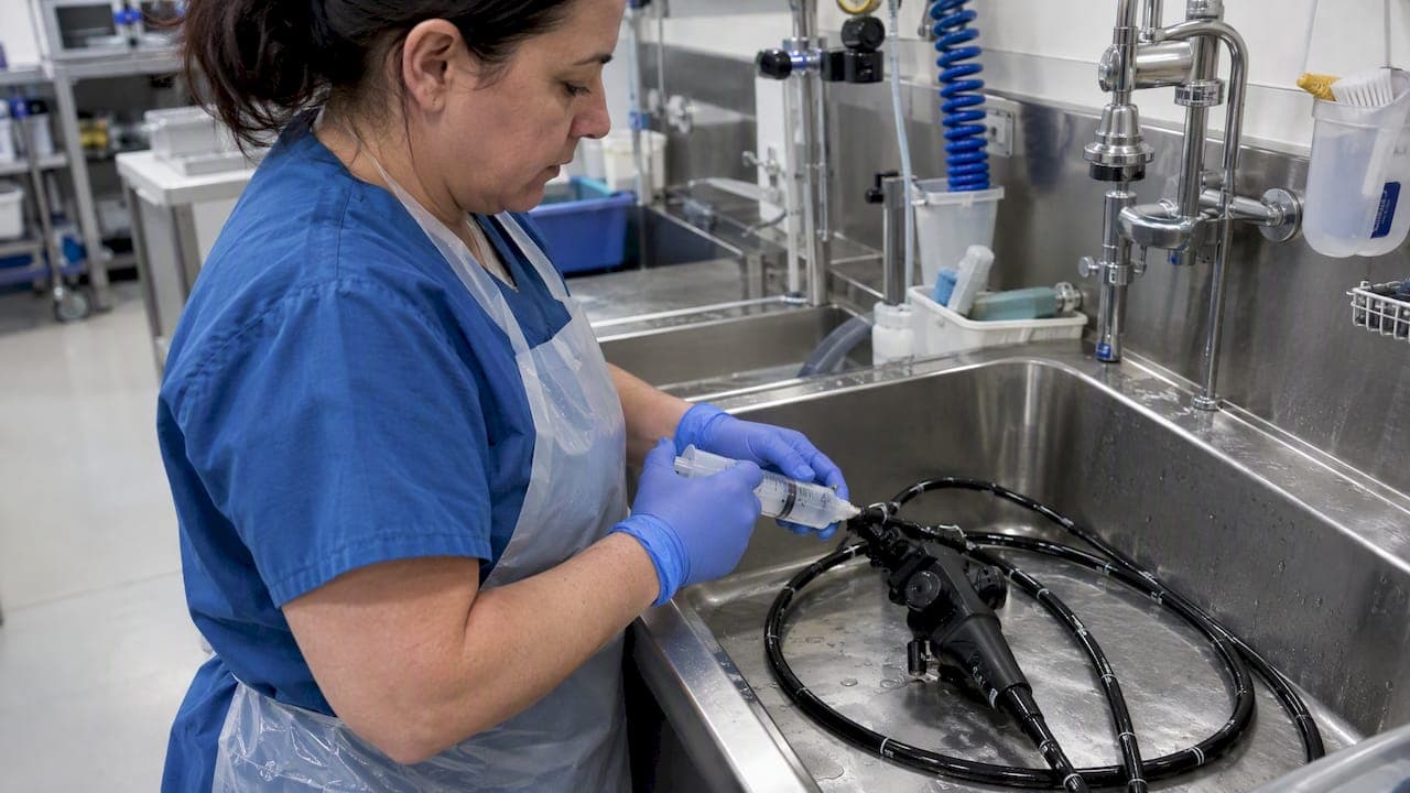
If a channel is clogged, start with controlled syringe flushing, then move to an enzymatic flush-brush-flush sequence before anything else. If residue persists after that, inspect the channel with a borescope and quarantine the scope rather than forcing another cleaning pass. Narrow, unbrushable channels retain residue more often than wider ones and sometimes need manufacturer evaluation before they go back into service.
TL;DR:
- Start precleaning within minutes of a procedure; dried blood, mucus, detergent, and simethicone can otherwise cling to channel walls.
- Match brush diameter to the channel: an undersized brush misses the wall, while an oversized one can bind or shed bristles inside the lumen.
- One randomized study found automated channel brushing lowered positive postcleaning cultures to 15.2%, versus 23.4% with manual brushing, supporting automation in high volume settings.
- Deep scratches, peeling, buckling, or perforation require quarantine and manufacturer evaluation, even when the channel still flushes freely after cleaning.
Table of Contents
- Why endoscope channels clog: anatomy, debris, and biofilm risk
- Immediate troubleshooting: safe first steps to clear a blocked channel
- Cleaning tools and techniques: brushes, syringes, detergents, and adjuncts
- Inspection and verification: borescopes and surveillance sampling for hidden blockages
- When to stop troubleshooting and send the scope for repair
- Prevention and program-level controls to reduce future clogs
- Research summary and practical takeaways
- What technicians consistently underestimate about channel care
- Tools and repair support for clogged channel problems
- FAQ
- Sources
Why endoscope channels clog: anatomy, debris, and biofilm risk
Not every channel in a flexible endoscope gets the same cleaning treatment, and that is exactly where clogs start. Air/water channels, auxiliary water channels, and elevator-wire channels on duodenoscopes are frequently too narrow for a brush to pass through, which means they rely almost entirely on flushing to stay clear. When flushing alone cannot physically scrub the lumen wall, anything that sticks tends to stay stuck.
The materials that cause blockages are rarely a mystery once you know what to look for:
- Tissue fragments and coagulated blood or mucus left behind after an incomplete precleaning step.
- Dried detergent residue from reprocessing cycles that ran too long between use and cleaning.
- Simethicone and other viscous anti-foaming agents, which are notorious for leaving a tacky film in narrow lumens.
- Broken accessory fragments, such as a biopsy forceps tip, that lodge in the channel and build debris around themselves.
Biofilm is what turns a simple residue problem into a persistent one. Once organic material adheres to a channel wall, bacteria can colonize it and form a protective matrix that passive flushing cannot dislodge. Borescope inspections have found that channel defects and retained debris are common across endoscope fleets, and that standard visual checks miss issues a scope exam catches.
During routine use, a few signs point to occlusion rather than a simple air/water malfunction: weak or absent suction, a water jet that sputters instead of flowing steadily, or resistance felt during syringe flushing at the end of a reprocessing cycle. Any of these warrants a closer look before the scope goes back into the schedule.
Immediate troubleshooting: safe first steps to clear a blocked channel
Start at the point of use. If a channel feels sluggish during a procedure or during immediate precleaning, do not wait for the full reprocessing cycle to address it. Catching resistance early prevents a soft blockage from drying into a hard one.
Pro Tip: Flush with the channel oriented the way the manufacturer's instructions for use (IFU) specify; flushing against the intended direction can push debris deeper into narrow segments instead of clearing it.
- Disconnect accessories and confirm the correct connector is attached for the channel you are flushing.
- Flush with a syringe of the size specified for that channel, using steady, moderate pressure rather than a hard push.
- If resistance remains, prepare an enzymatic detergent solution per the IFU and allow the recommended soak time before repeating the flush.
- Follow the soak with a flush-brush-flush sequence where the channel is brushable, using a brush sized to the lumen.
- For channels too narrow to brush, repeat low-pressure flushing or backflushing once; avoid pulsatile high-pressure attempts beyond what the IFU allows, since excessive force can abrade or perforate the channel wall.
- If the obstruction has not cleared after one full repeat cycle, stop. Tag the scope for inspection instead of attempting a third round.
The decision point at step six matters more than it looks. Repeated forceful attempts to push a blockage through are a common way technicians accidentally convert a cleanable residue problem into a structural defect, which is harder and more expensive to fix than the original clog.
Cleaning tools and techniques: brushes, syringes, detergents, and adjuncts
Brush selection is not a minor detail. A brush that is undersized will not contact the channel wall, and one that is oversized can bind and tear bristles loose inside the lumen. Matching brush diameter to the specific channel, whether it is a biopsy channel, suction channel, or auxiliary line, is the difference between a thorough clean and a false sense of one. Our guide on sizing endoscope brushes to channel lumen diameters covers the sizing logic in more detail.
A few habits protect both the scope and the cleaning outcome:
- Inspect every brush after each pass for missing or bent bristles, since a shedding brush can leave fragments inside the channel.
- Retire single-use brushes after one use and inspect reusable brushes under good lighting before reuse.
- Match syringe size to channel diameter so pressure stays within the range the IFU allows.
- Use connectors rated for the specific channel port rather than improvising a fit.
Flush-brush-flush, or FBF, is the technique that actually resolves what passive flushing leaves behind. CDC guidance on gastrointestinal endoscope reprocessing notes that manual flushing alone often fails to clear narrow, unbrushable channels, and that FBF with friction recovers substantially more residue than flushing by itself. The same guidance outlines FBF as the basis for duodenoscope surveillance sampling protocols developed with the FDA and the American Society for Microbiology.
One randomized study found that automated channel brushing reduced positive post-cleaning cultures to 15.2%, compared with 23.4% for manual brushing. That gap suggests automation can meaningfully reduce residual bioburden in settings where staffing or throughput makes consistent manual technique hard to guarantee.
On the chemical side, enzymatic cleaners remain the standard for breaking down protein-based residue, and detergent compatibility with channel materials should always follow the IFU rather than a generalized assumption. Plasma-activated disinfectants and non-thermal plasma technologies are showing strong results against biofilm in lab settings, but they remain investigational and should not substitute for IFU-compliant reprocessing today.
Inspection and verification: borescopes and surveillance sampling for hidden blockages

A borescope exam is warranted whenever an obstruction persists after one full troubleshooting cycle, as part of a scheduled surveillance program, or when a technician suspects internal damage from a prior forceful flush. Visual inspection of the exterior tells you almost nothing about what is happening inside a 2 mm lumen.
The technique follows a consistent path:
- Prepare the borescope per its own disinfection protocol before and after each use to avoid cross-contaminating the channel under inspection.
- Advance from the channel inlet through the shaft toward the distal tip, documenting findings at each segment.
- Look for discoloration, pitting, peeling, retained debris, or any narrowing that suggests built-up residue rather than a clear lumen.
- Photograph or record findings so severity can be tracked over time rather than relying on memory.
A simple severity framework helps standardize the decision that follows an exam: light, loose debris that clears with another FBF cycle; adherent residue or discoloration that warrants a repeat inspection after additional cleaning; and structural findings such as scratches or peeling that call for manufacturer evaluation regardless of how the channel flushes afterward. Surveillance culturing, using FBF-based sampling with ATP or microbial culture testing, provides the quality assurance layer that confirms whether cleaning protocols are actually working, rather than assuming they are because the channel flushes freely.
When to stop troubleshooting and send the scope for repair
Some findings mean the cleaning conversation is over and the repair conversation starts. A borescope exam that shows a deep scratch, visible peeling, buckling of the inner lining, or any perforation is a stop signal, not a cue for another cleaning attempt. Adherent residue that reappears after a full FBF cycle and a follow-up inspection falls into the same category.
- Deep scratches, peeling, buckling, or perforation seen on borescope exam.
- Residue that remains adherent after a complete flush-brush-flush cycle and re-inspection.
- Repeated surveillance-culture failures on the same device despite correct reprocessing technique.
- Any uncertainty about whether a retained accessory fragment has been fully removed.
Once a scope meets any of these criteria, quarantine it immediately, label it clearly as out of service, and document the finding with photos or borescope footage. Chain-of-custody records matter here: note who identified the issue, when, and what steps preceded the decision to pull the device, since that record supports both infection control review and the manufacturer's repair intake process. Following the manufacturer's stated timelines for repair requests, as set out in the device IFU, keeps the return process from adding unnecessary downtime.
Prevention and program-level controls to reduce future clogs
Most clogged channels trace back to a gap in one of the seven essential reprocessing steps: point-of-use precleaning, leak testing, manual cleaning with brushing and flushing, visual inspection, high-level disinfection or sterilization, rinsing, and drying and storage. CDC guidance frames these as a connected sequence, and skipping or rushing any one of them raises the odds that residue hardens before the next step can remove it.
Pro Tip: Time-to-precleaning matters more than most programs treat it: start precleaning within minutes of the procedure ending, before organic material has a chance to dry inside the channel.
- Precleaned immediately after use, every time, with no exceptions for a busy schedule.
- Minimize simethicone and other viscous lubricants where clinically appropriate, since they are a frequent cause of channel residue.
- Train staff on brush sizing, flush technique, and borescope use, then audit adherence to the written IFU on a recurring basis.
- Dry channels fully with filtered, forced air before storage, and store scopes vertically in a cabinet designed to prevent recontamination.
- Set a borescope surveillance schedule based on usage volume and channel complexity rather than inspecting only when a problem is already suspected.
Automated channel-brushing systems are worth considering for programs with high case volume, given the randomized evidence that they reduce positive culture rates compared with manual brushing alone. For reference on the drying and storage steps that close out this sequence, our walkthrough of endoscope sterilization workflows covers the practical sequencing in more detail.
Research summary and practical takeaways
The evidence behind these recommendations is consistent across several independent lines of research. CDC and FDA guidance endorses flush-brush-flush as the basis for surveillance sampling because it recovers more residue than passive flushing, particularly in channels too narrow to brush directly.

A randomized study found automated channel brushing cut positive post-cleaning cultures to 15.2%, against 23.4% with manual brushing alone, a gap that argues for automation in high-volume settings.
Borescope studies add another layer: exams have found channel defects and retained debris to be common findings across inspected fleets, and a deep learning model applied to borescope video achieved sensitivity above 90% for detecting any channel abnormality, suggesting routine inspection could become far more scalable. On the chemistry side, non-thermal plasma technology has eliminated biofilm completely in laboratory surrogate channels, but the authors themselves call for further real-world validation before it replaces standard reprocessing.
What technicians consistently underestimate about channel care
The biggest gap we see is not a lack of protocols. Most programs have an IFU, a cleaning log, and a flushing procedure on paper. The gap is in the few minutes right after a procedure ends, when precleaning either happens immediately or gets pushed back by a busy room turnover. That delay is where residue sets and biofilm gets its start.
Investing in the right small-diameter brushes, properly sized syringes, and a borescope for routine checks pays off before a channel ever reaches the point of a hard clog. Technicians can find these tools and repair services to help close that gap in their own programs.
— Endoscope
Tools and repair support for clogged channel problems
We stock the inspection and cleaning hardware that makes the troubleshooting steps above practical day to day, not just theoretical. When a channel will not clear with standard flushing, having the right borescope and brush on hand turns a guessing game into a documented decision.

- Inspection borescopes and videoscopes sized for routine channel surveillance and persistent-blockage checks.
- Channel brushes matched to lumen diameter, built to reduce the risk of bristle shedding during an FBF cycle.
- Flexible biopsy forceps and compatible accessories designed to reduce the risk of fragment retention in the first place.
- Endoscope repair services for scopes that fail borescope inspection or repeated surveillance culturing.
Browse our medical endoscopes and accessories to find the tools your reprocessing team needs, or reach out through our repair services page to arrange a manufacturer evaluation for a scope you have already quarantined.
FAQ
What is the working channel of an endoscope?
The working channel is the internal lumen that allows instruments, such as biopsy forceps, suction, and irrigation fluid, to pass through the length of the endoscope to the distal tip. Its narrow diameter, often too small for a brush in auxiliary or elevator-wire channels, is exactly why it is prone to retain debris.
Can I have an endoscopy with a blocked nose?
This question concerns a clinical procedure decision for the patient and the performing physician, not a device-reprocessing issue, so it falls outside device maintenance guidance. A physician's office can advise whether a scheduled procedure needs to be rescheduled based on the specific type of endoscopy and the patient's symptoms.
How do I properly clean an endoscope channel?
Correct cleaning follows the seven essential reprocessing steps: precleaning, leak testing, manual cleaning with flush-brush-flush, visual inspection, high-level disinfection or sterilization, rinsing, and drying and storage, as outlined in CDC guidance. Flush-brush-flush recovers more residue than flushing alone, especially in narrow channels that cannot be brushed directly.
What is the risk of infection after an endoscopy?
Infection risk after endoscopy is tied closely to reprocessing quality, since retained debris and biofilm in channels have been linked to documented transmission events in outbreak investigations. Programs that follow complete reprocessing steps and use borescope surveillance to catch hidden defects reduce that risk, though no reprocessing protocol eliminates it entirely.
What should I check before deciding to continue cleaning a blocked channel myself?
Check whether the blockage has cleared after one full flush-brush-flush cycle and whether a borescope exam shows only loose debris versus adherent residue or structural damage. If damage such as scratching, peeling, or buckling is visible, or if resistance persists after a complete repeat cycle, the scope should be quarantined for manufacturer evaluation rather than cleaned again.
Sources
- Borescope inspection of endoscope working channels: Why and how?
- Effect of an automated flexible endoscope channel brushing system on improving reprocessing quality
- Gastrointestinal flexible endoscopes: infection control risks, lessons learned from outbreaks, and CDC guidance
- Direct application of non-thermal plasma technology for the elimination of biofilm from endoscope channels